

Observer reliability of three-dimensional cephalometric landmark identification on cone-beam computerized tomography
- PMID: 18718796
- PMCID: PMC2642991
- DOI: 10.1016/j.tripleo.2008.05.039
Observer reliability of three-dimensional cephalometric landmark identification on cone-beam computerized tomography
Abstract
Objective: To evaluate reliability in 3-dimensional (3D) landmark identification using cone-beam computerized tomography (CBCT).
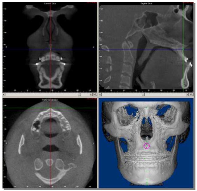
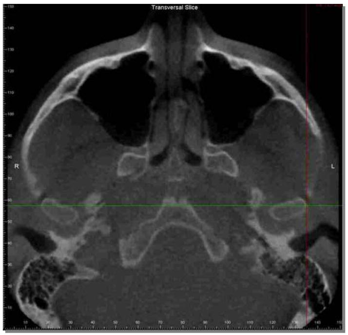
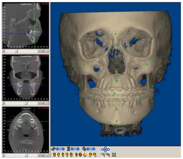
Study design: Twelve presurgery CBCTs were randomly selected from 159 orthognathic surgery patients. Three observers independently repeated 3 times the identification of 30 landmarks in the sagittal, coronal, and axial slices. A mixed-effects analysis of variance model estimated the intraclass correlations (ICC) and assessed systematic bias.
Results: The ICC was >0.9 for 86% of intraobserver assessments and 66% of interobserver assessments. Only 1% of intraobserver and 3% of interobserver coefficients were <0.45. The systematic difference among observers was greater in X and Z than in Y dimensions, but the maximum mean difference was quite small.
Conclusion: Overall, the intra- and interobserver reliability was excellent. Three-dimensional landmark identification using CBCT can offer consistent and reproducible data if a protocol for operator training and calibration is followed. This is particularly important for landmarks not easily specified in all 3 planes of space.
Figures
References
-
- Kumar V, Ludlow JB, Mol A, Cevidanes L. Comparison of conventional and cone beam CT synthesized cephalograms. Dentomaxillofac Radiol. 2007;36:263–9. - PubMed
-
- Halazonetis DJ. From 2-dimensional cephalograms to 3-dimensional computed tomography scans. Am J Orthod Dentofacial Orthop. 2005;127:627–637. - PubMed
-
- Lou L, Lagravere MO, Compton S, Major PW, Flores-Mir C. Accuracy of measurements and reliability of landmark identification with computed tomography (CT) techniques in the maxillofacial area: a systematic review. Oral Surg Oral Med Oral Pathol Oral Radiol Endod. 2007;104:402–11. - PubMed
-
- Park SH, Yu HS, Kim KD, Lee KJ, Baik HS. A proposal for a new analysis of craniofacial morphology by 3-dimensional computed tomography. Am J Orthod Dentofacial Orthop. 2006;129:600, e23–34. - PubMed
-
- Scarfe WC, Farman AG, Sukovic P. Clinical applications of cone-beam computed tomography in dental practice. J Can Dent Assoc. 2006;72:75–80. - PubMed
Publication types
MeSH terms
Grants and funding
LinkOut - more resources
Full Text Sources
Other Literature Sources
Medical
