Review
. 2008 Nov 1;423(2):97-107.
doi: 10.1016/j.gene.2008.07.015. Epub 2008 Jul 22.

Alternative splicing in the NF-kappaB signaling pathway

Affiliations
Review

Alternative splicing in the NF-kappaB signaling pathway

Joshua R Leeman et al. Gene. .

Abstract

Activation of transcription factor NF-kappaB can affect the expression of several hundred genes, many of which are involved in inflammation and immunity. The proper NF-kappaB transcriptional response is primarily regulated by post-translational modification of NF-kappaB signaling constituents. Herein, we review the accumulating evidence suggesting that alternative splicing of NF-kappaB signaling components is another means of controlling NF-kappaB signaling. Several alternative splicing events in both the tumor necrosis factor and Toll/interleukin-1 NF-kappaB signaling pathways can inhibit the NF-kappaB response, whereas others enhance NF-kappaB signaling. Alternative splicing of mRNAs encoding some NF-kappaB signaling components can be induced by prolonged exposure to an NF-kappaB-activating signal, such as lipopolysaccharide, suggesting a mechanism for negative feedback to dampen excessive NF-kappaB signaling. Moreover, some NF-kappaB alternative splicing events appear to be specific for certain diseases, and could serve as therapeutic targets or biomarkers.


Figures
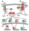
Fig. 1
Fig. 1. Shown are the TNFR and TLR/IL-1R (TIR) signaling pathways leading to activation of NF-κB
(A) Following TNF binding to the TNFR, the adaptor proteins TRADD, TRAF2, and RIP are recruited to the receptor. RIP mediates activation of the IKK complex through ubiquitin-dependent interaction with NEMO. (CYLD and A20 are deubiquitiases that can remove K63 ubiqutin from the indicated proteins to arrest NF-κB activation.) (B) Binding of LPS or IL-1 results in receptor dimerization and recruitment of the adaptor proteins TRIF, MyD88 and TIRAP. This receptor complex then recruits IRAK1, which becomes activated by IRAK4. Activated IRAK1 leaves the receptor complex and binds TRAF6. TAB2 then mediates interaction of activated TRAF6 with the TAK1/TAB1 complex. TAK1 activates the IKK complex by phosphorylation of IKKβ. (C) Activated IKK phosphorylates IκBα or IκBβ, which leads to their degradation and thus nuclear localization of NF-κB transcription factor dimers. In the alternative NF-κB pathway (not depicted), activated IKKα phosphorylates p100 (or p105), targeting it for processing by the proteasome into the mature p52 (or p50) protein. Dimers containing these processed p52 or p50 proteins can then enter the nucleus. Shown in green are NF-κB signaling components that can be alternatively spliced to enhance NF-κB signaling; in red are those where alterative splicing can inhibit the NF-κB response; ones in green and red have both inhibitory and activating splice variants; in white are NF-κB signaling proteins where alternative splicing is not yet known to affect the NF-κB response. Arrows indicate activating steps; double-sided arrows indicate reversible interactions; and bars (as with CYLD and A20) indicate inhibitory effects.
Fig. 2
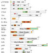
Fig. 2. Structural effects of alternative splicing on NF-κB signaling components that can positively or negatively affect NF-κB signaling
Shown are schematics of alternatively spliced NF-κB signaling proteins. Proteins and protein domains are outlined in black. Numbering above protein structures indicate aa numbers. Alternative splicing events resulting in deletions are outlined in yellow. Horizontal yellow in the center of a box indicate overlapping alternative splicing events in the same protein. Alternative splicing insertions are denoted by inverse triangles. The dotted line (in Tlr4) designates a stop codon that is caused by alternative splicing. Alternative splicing events that enhance NF-κB signaling are shown in green; alternative splicing events that inhibit NF-κB are shown in red (see also Fig. 1). Abbreviations: BD, TRAF2/NEMO/Bcl-3 binding domain; CC1, coiled-coil domain 1; CC2, coiled-coil domain 2; CD, cytoplasmic domain; DD, death domain; DUB, deubiquitinase domain; ED, extracellular domain; ID, intermediate domain; LRR, leucine-rich region; LZ, leucine zipper; RHD, REL homology domain; RID, REL inhibitory domain; RF, ring finger; TAD, transactivation domain; TD, TRAF domain; TIR, Toll/IL-1 interaction domain; TM, transmembrane domain; ZF, zinc finger.

References

    1. Arend WP. The balance between IL-1 and IL-1Ra in disease. Cytokine Growth Factor Rev. 2002;13:323–340. - PubMed
    1. Arend WP, Guthridge CJ. Biological role of interleukin 1 receptor antagonist isoforms. Ann. Rheum. Dis. 2000;59:60–64. - PMC - PubMed
    1. Beg AA, Baltimore D. An essential role for NF-κB in preventing TNF-α-induced cell death. Science. 1996;274:782–784. - PubMed
    1. Beg AA, Sha WC, Bronson RT, Ghosh S, Baltimore D. Embryonic lethality and liver degeneration in mice lacking the RelA component of NF-κB. Nature. 1995;376:167–170. - PubMed
    1. Bignell GR, et al. Identification of the familial cylindromatosis tumour-suppressor gene. Nat. Genet. 2000;25:160–165. - PubMed

Publication types

LinkOut - more resources