. 2008 Nov 21;283(47):32802-11.
doi: 10.1074/jbc.M803440200. Epub 2008 Aug 20.

Follistatin-like 1, a secreted muscle protein, promotes endothelial cell function and revascularization in ischemic tissue through a nitric-oxide synthase-dependent mechanism

Affiliations

Follistatin-like 1, a secreted muscle protein, promotes endothelial cell function and revascularization in ischemic tissue through a nitric-oxide synthase-dependent mechanism

Noriyuki Ouchi et al. J Biol Chem. .

Abstract

Myogenic Akt signaling coordinates blood vessel recruitment with normal tissue growth. Here, we investigated the role of Follistatin-like 1 (Fstl1) in the regulation of endothelial cell function and blood vessel growth in muscle. Transgenic Akt1 overexpression in skeletal muscle led to myofiber growth that was coupled to an increase in muscle capillary density. Myogenic Akt signaling or ischemic hind limb surgery led to the induction of Fstl1 in muscle and increased circulating levels of Fstl1. Intramuscular administration of an adenoviral vector expressing Fstl1 (Ad-Fstl1) accelerated flow recovery and increased capillary density in the ischemic hind limbs of wild-type mice, and this was associated with an increase in endothelial nitric oxide synthase (eNOS) phosphorylation at residue Ser-1179. In cultured endothelial cells, Ad-Fstl1 stimulated migration and differentiation into network structures and inhibited apoptosis under conditions of serum deprivation. These cell responses were associated with the activating phosphorylation of Akt and eNOS. Conversely, transduction with dominant-negative Akt or LY294002 blocked Fstl1-stimulated eNOS phosphorylation and inhibited Fstl1-stimulated cellular responses. Treatment with the eNOS inhibitor N(G)-nitro-L-arginine methyl ester also reduced endothelial cell migration and differentiation induced by Ad-Fstl1. The stimulatory effect of Ad-Fstl1 on ischemic limb reperfusion was abolished in mice lacking eNOS. These data indicate that Fstl1 is a secreted muscle protein or myokine that can function to promote endothelial cell function and stimulates revascularization in response to ischemic insult through its ability to activate Akt-eNOS signaling.


Figures

FIGURE 1.
FIGURE 1.
Transgenic Akt activation increases capillary density and Fstl1 expression in gastrocnemius muscle. A, increased capillary vessels in gastrocnemius muscles in muscle-specific Akt-TG mice following 2 weeks of Akt1 activation. Immunostaining of gastrocnemius tissues of control (n = 5) and Akt-TG (n = 5) mice was performed with anti-CD31 monoclonal antibody. Capillary density was expressed as the number of capillaries per high power field. Results are shown as the mean ± S.E. *, p < 0.01 versus control mice. B, up-regulation of Fstl1 expression in gastrocnemius muscle and serum in muscle-specific Akt-TG mice after Akt1 activation for 2 weeks. Fstl1 expression was determined by QRT-PCR and Western blot analyses (n = 5). Fstl1 mRNA levels were expressed relative to levels of glyceraldehyde-3-phosphate dehydrogenase mRNA. Results are expressed relative to control. Relative protein levels of Fstl1 were quantified (n = 4-5) by using ImageJ. Results are shown as the mean ± S.E. *, p < 0.01 versus control. C, Fstl1 is secreted from C2C12 myotube cultures. C2C12 cells were transduced with adenoviral vectors expressing Fstl1 (Ad-Fstl1), or β-galactosidase (Ad-β-gal) for 16 h followed by 24 h of incubation in serum-free media. Fstl1 protein levels were determined in media, and cell lysates were determined by Western blot analysis. Representative blots are shown from three independent experiments.
FIGURE 2.
FIGURE 2.
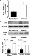
Elevated Fstl1 levels in ischemic muscle and serum after hind limb ischemic surgery. A, Fstl1 expression in non-ischemic or ischemic adductor muscles was measured by QRT-PCR (n = 5) at day 7 after femoral artery resection. Fstl1 transcript levels were expressed relative to levels of glyceraldehyde-3-phosphate dehydrogenase mRNA. Results are expressed relative to control. B, Fstl1 expression in non-ischemic (n = 4) and ischemic skeletal muscle (n = 4) was measured by Western blot analyses on the postoperative day 14. Serum was collected from control (n = 4) or mice subjected to hind limb ischemic surgery (n = 4), and Fstl1 levels were determined by Western blot analyses. Relative protein levels of Fstl1 were quantified (n = 4) by using ImageJ. Results are shown as the mean ± S.E. *, p < 0.05 versus non-ischemic.
FIGURE 3.
FIGURE 3.
Fstl1 promotes perfusion recovery and capillary vessel formation of ischemic limbs in mice in vivo. Adenoviral vectors expressing Fstl1 (Ad-Fstl1), or β-galactosidase (Ad-β-gal, control) were injected into five sites in adductor muscle of wild-type mice (2 × 10⁸ pfu each) at 3 days prior to ischemic surgery. A, Fstl1 expression in ischemic muscle at 6 days after injection of Ad-Fstl1 or Ad-β-gal. Fstl1 protein expression was determined by Western blot analysis. Representative blots are shown from five independent experiments. B, quantitative analysis of the ischemic/non-ischemic laser Doppler blood flow ratio in wild-type mice treated with Ad-Fstl1 (n = 8) and Ad-β-gal (n = 8). Results are shown as the mean ± S.D. *, p < 0.05 versus control mice. **, p < 0.01 versus control mice. C, quantitative analysis of capillary density in ischemic muscles of wild-type mice treated with Ad-Fstl1 (n = 5) and Ad-β-gal (n = 5) on postoperative day 14. Immunostaining of ischemic tissues was performed with anti-CD31 monoclonal antibody. Capillary density was expressed as the number of capillaries per muscle fiber. Results are shown as the mean ± S.E. *, p < 0.01 versus control mice. D, quantitative analysis of capillary density in non-ischemic gastrocnemius muscles of wild-type mice treated with Ad-Fstl1 (n = 4) and Ad-β-gal (n = 4) at day 7 after injection (2 × 10⁸ pfu each). Immunostaining of gastrocnemius muscle tissues was performed with anti-CD31 monoclonal antibody. Capillary density was expressed as the number of capillaries per muscle fiber. Results are shown as the mean ± S.E.
FIGURE 4.
FIGURE 4.
Fstl1 promotes endothelial cell migration and differentiation into vascular-like structures. A, expression of Fstl1 protein in media and cell lysates from HUVECs. HUVECs were transduced with Ad-Fstl1 and Ad-β-gal for 8 h followed by 24 h of incubation in serum-free media. Fstl1 protein levels were determined in media, and cell lysates were determined from HUVECs by Western blot analysis. Representative blots are shown from four independent experiments. B, endothelial cell network formation in response to Fstl1. After 24 h of serum deprivation and transduction with Ad-Fstl1 and Ad-β-gal, HUVECs were seeded on Matrigel-coated culture dishes. Representative cultures are shown (upper panel). Quantitative analyses of network formation are shown (bottom panel). C, migratory activities of HUVECs following treatment with Fstl1. A modified Boyden chamber assay was performed using HUVECs transduced with Ad-Fstl1 and Ad-β-gal. Results are shown as the mean ± S.E. (n = 7-8). Results are expressed relative to the values compared with control. *, p < 0.01 versus Ad-β-gal.
FIGURE 5.
FIGURE 5.
Fstl1 protects endothelial cells from apoptosis. HUVECs were transduced with Ad-Fstl1 and Ad-β-gal for 8 h followed by incubation with serum-free media for 48 h. A, inhibitory effect of Fstl1 on nucleosome fragmentation of HUVECs. Nucleosome fragmentation was assessed by enzyme-linked immunosorbent assay. Results are expressed relative to the values compared with control. B, inhibition of HUVEC death by Fstl1 assessed by a quantitative MTS-based assay. C, the frequency of TUNEL-positive HUVECs is reduced after treatment with Fstl1. Representative photomicrographs of TUNEL-positive HUVECs are shown (upper panels). Quantitative analyses of the frequency of TUNEL-positive HUVECs are shown (bottom panel). Apoptotic nuclei were identified by TUNEL staining (green), and total nuclei were identified by 4′,6-diamidino-2-phenylindole counterstaining (blue). Results are shown as the mean ± S.E. (n = 8-10). *, p < 0.01 versus Ad-β-gal.
FIGURE 6.
FIGURE 6.
Fstl1-stimulated endothelial cell responses are dependent on Akt signaling. A, Fstl1-stimulated signaling in endothelial cells. HUVECs were transduced with Ad-Fstl1 and Ad-β-gal for 8 h followed by 24 h of incubation with serum-free media. Changes in the phosphorylation of eNOS (P-eNOS), Akt (P-Akt), GSK (P-GSK), and ERK (P-ERK) following Ad-Fstl1 treatment were determined by Western blot analysis. Representative blots are shown. Relative phosphorylation levels of eNOS and Akt were quantified (n = 6) by using ImageJ. Immunoblots were normalized to total loaded protein. B, role of Akt in regulation of Fstl1-induced signaling. HUVECs were infected with adenoviral constructs encoding dominant-negative Akt1 (Ad-dnAkt) or Ad-β-gal at an m.o.i. of 10 along with Ad-Fstl1 or Ad-β-gal at an m.o.i. of 10 for 8 h, followed by serum deprivation for 24 h. Phosphorylation of eNOS (P-eNOS) and Akt (P-Akt) were determined by Western blot analysis. Representative blots are shown from four independent experiments. C and D, contribution of Akt to Fstl1-mediated cellular responses. HUVECs were transduced with Ad-dnAkt or Ad-β-gal along with Ad-Fstl1 or Ad-β-gal for 8 h. After 24 h of serum deprivation, Matrigel (C) or modified Boyden chamber assays (D) were performed. E, involvement of Akt in Fstl1-induced endothelial cell survival. After transduction with Ad-dnAkt or Ad-β-gal along with Ad-Fstl1 or Ad-β-gal for 8 h, cells were incubated in serum-free media. Nucleosome fragmentation was assessed by enzyme-linked immunosorbent assay. Results are shown as the mean ± S.E. (n = 6-8). Results are expressed relative to the values compared with control. *, p < 0.01.
FIGURE 7.
FIGURE 7.
PI3K and eNOS signaling is involved in Fstl1-induced endothelial cell responses. A, effect of LY294002 on Fstl1-induced phosphorylation of eNOS and Akt. HUVECs were treated with LY294002 (10 μm) or vehicle following transduction with Ad-Fstl1 or Ad-β-gal. After 24 h serum deprivation, phosphorylation of eNOS (P-eNOS), and Akt (P-Akt) were determined by Western blot analysis. Representative blots are shown. Relative phosphorylation levels of eNOS and Akt were quantified (n = 4) by using ImageJ. Immunoblots were normalized to total loaded protein. B and C, contribution of PI3K to Fstl1-mediated endothelial differentiation and migration. HUVECs were treated with LY294002 (10 μm), l-NAME (1 mg/ml), or vehicle along with Ad-Fstl1 or Ad-β-gal for 8 h. After 24-h serum-starvation, Matrigel (B) or modified Boyden chamber assays (C) were performed. Results are shown as the mean ± S.E. (n = 5-8). Results are expressed relative to the values compared with control. *, p < 0.01.
FIGURE 8.
FIGURE 8.
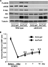
Fstl1 stimulates ischemia-induced revascularization through an eNOS-dependent mechanism. A, phosphorylation of eNOS and Akt in ischemic muscle tissues of wild-type and eNOS-KO mice at 6 days after transduction with Ad-Fstl1 or Ad-β-gal. Ad-Fstl1 or Ad-β-gal (control) was injected into five sites in adductor muscle of wild-type and eNOS-KO mice (2 × 10⁸ pfu each), 3 days before ischemic surgery. Phosphorylation of eNOS (P-eNOS) and Akt (P-Akt), total eNOS, total Akt, and Fstl1 levels were analyzed by Western blotting. Representative blots are shown from four independent experiments. B, quantitative analysis of the ischemic/non-ischemic laser Doppler blood flow ratio in eNOS-KO mice treated with Ad-Fstl1 (n = 7) and Ad-β-gal (n = 7). Results are shown as the mean ± S.D. N.S., not significant.

Similar articles

Cited by

References

    1. Degens, H., Veerkamp, J. H., van Moerkerk, H. T. B., Turek, Z., Hoofd, L. J. C., and Binkhorst, R. A. (1993) Int. J. Biochem. 25 1141-1148 - PubMed
    1. Degens, H., Turek, Z., Hoofd, L. J. C., Van't Hof, M. A., and Binkhorst, R. A. (1992) J. Anat. 180 455-463 - PMC - PubMed
    1. Ingjer, F. (1979) Eur. J. Appl. Physiol. Occup. Physiol. 40 197-209 - PubMed
    1. Kano, Y., Shimegi, S., Masuda, K., Ohmori, H., and Katsuta, S. (1997) Eur. J. Appl. Physiol. Occup. Physiol. 75 97-101 - PubMed
    1. Shiojima, I., and Walsh, K. (2006) Genes Dev. 20 3347-3365 - PubMed

Publication types

Substances

LinkOut - more resources