. 2008 Oct 24;283(43):29447-60.
doi: 10.1074/jbc.M804595200. Epub 2008 Aug 21.

Targeted induction of lung endothelial cell apoptosis causes emphysema-like changes in the mouse

Affiliations

Targeted induction of lung endothelial cell apoptosis causes emphysema-like changes in the mouse

Ricardo J Giordano et al. J Biol Chem. .

Abstract

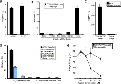
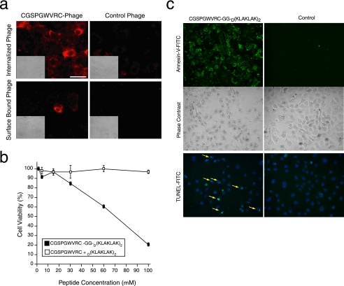
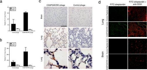
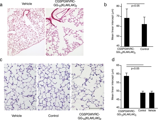
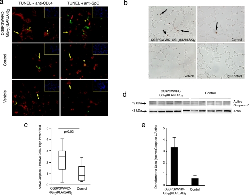
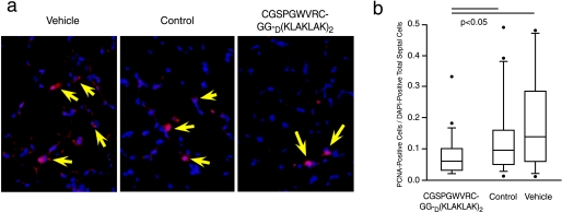
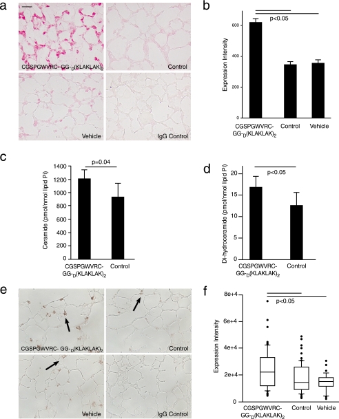
Pulmonary gas exchange relies on a rich capillary network, which, together with alveolar epithelial type I and II cells, form alveolar septa, the functional units in the lung. Alveolar capillary endothelial cells are critical in maintaining alveolar structure, because disruption of endothelial cell integrity underlies several lung diseases. Here we show that targeted ablation of lung capillary endothelial cells recapitulates the cellular events involved in cigarette smoke-induced emphysema, one of the most prevalent nonneoplastic lung diseases. Based on phage library screening on an immortalized lung endothelial cell line, we identified a lung endothelial cell-binding peptide, which preferentially homes to lung blood vessels. This peptide fused to a proapoptotic motif specifically induced programmed cell death of lung endothelial cells in vitro as well as targeted apoptosis of the lung microcirculation in vivo. As early as 4 days following peptide administration, mice developed air space enlargement associated with enhanced oxidative stress, influx of macrophages, and up-regulation of ceramide. Given that these are all critical elements of the corresponding human emphysema caused by cigarette smoke, these data provide evidence for a central role for the alveolar endothelial cells in the maintenance of lung structure and of endothelial cell apoptosis in the pathogenesis of emphysema-like changes. Thus, our data enable the generation of a convenient mouse model of human emphysema. Finally, combinatorial screenings on immortalized cells followed by in vivo targeting establishes an experimental framework for discovery and validation of additional ligand-directed pharmacodelivery systems.


Figures

FIGURE 1.
FIGURE 1.
CGSPGWVRC phage binds specifically to lung endothelial cells. a, CGSPGWVRC phage binding to primary mouse lung microvascular endothelial cells cultured at either 33 or 37 °C detected by the BRASIL method. b, CGSPGWVRC phage binds specifically to lung endothelial cells but not to endothelial cells derived from control organs (brain, prostate, bone marrow, or kidney). c, CGSPGWVRC phage binding ex vivo to single-cell suspensions prepared from mouse lungs. CGSPGWVRC phage did not bind to single-cell suspensions prepared from the control organ (spleen). d, mutation of certain residues within CGSPGWVRC (to alanine) abolishes phage binding to lung endothelial cells. Bone marrow endothelial cells served as the negative control. Mutant residues are color-coded. e, CGSPGWVRC phage binding to lung endothelial cells is inhibited in a dose-dependent manner by the cognate synthetic peptide. An unrelated cyclic control peptide did not affect the phage binding. Shown are means ± S.E. from triplicate samples. Insertless control phage served as a negative control phage in a-d. Fd-tet phage values were set to 1 in a-c.
FIGURE 2.
FIGURE 2.
CGSPGWVRC peptide mediates internalization of ligands into lung endothelial cells and cell apoptosis. a, CGSPGWVRC peptide mediates phage internalization into lung endothelial cells. CGSPGWVRC phage or control phage were incubated with lung endothelial cells at 37 °C to allow for phage internalization. Cells were stained with an anti-bacteriophage antibody after removal of the membrane-bound phage. The upper panels show staining of permeabilized cells revealing the internalized phage. The lower panels show staining of nonpermeabilized cells, demonstrating the successful removal of phage from the cell surface. Scale bar, 100 μm. b, lung endothelial cells were incubated with increasing concentrations (up to 100 μm) of proapoptotic peptide synthesized in conjunction with the targeting CGSPGWVRC peptide (CGSPGWVRC-GG-D(KLAKLAK)2) or negative control peptides (an equimolar mixture of CGSPGWVRC and D(KLAKLAK)2). Cell viability was determined by optical absorbance using a cell proliferation detection reagent. Shown are means ± S.E. from triplicate wells. c, lung endothelial cells were incubated with 100 μm proapoptotic peptide CGSPGWVRC-GG-D(KLAKLAK)2 or negative control peptides (equimolar mixture of CGSPGWVRC and D(KLAKLAK)2) for 2 h. Induction of cell apoptosis was detected by Annexin-V-fluorescein isothiocyanate (FITC) binding (top and middle panels) or TUNEL (bottom, yellow arrows).
FIGURE 3.
FIGURE 3.
Targeting mouse lung vasculature in vivo. a, the ability of the CGSPGWVRC phage to home to mouse lungs was evaluated after intravenous phage administration into BALB/c mice. Phage were recovered 10 min later from lungs or control tissues after perfusion. Shown are mean ± S.E. of TU from three mice and triplicate plating. b, the ability of the CGSPGWVRC phage to home and internalize to mouse lungs was evaluated 24 h after an intravenous phage administration into BALB/c mice. Phage were recovered from lungs or control tissues without perfusion. Shown are mean ± S.E. of TU from three mice and triplicate plating. c, CGSPGWVRC phage or control phage were administered intravenously into mice. Mice were perfused 10 min after the phage injections, and lungs and control organ were recovered. A bacteriophage-specific antibody was used for staining. Scale bar, 100 μm for the panels in the top and middle rows and 20 μm for the bottom row. Insertless phage served as a negative control. d, targeting of CGSPGWVRC synthetic peptide to the lung vasculature was evaluated by intravenous administration of CGSPGWVRC-biotin. After 1 h of circulation, peptide was detected using a streptavidin-fluorescein isothiocyanate conjugate (yellow) and co-stained with an endothelial marker specific antibody (CD31; red). Vascular co-localization of CGSPGWVRC-biotin is indicated by the yellow areas in the merged image. No specific homing was observed to a control organ or when a control peptide-biotin was injected into mice.
FIGURE 4.
FIGURE 4.
Induction of morphological changes in mouse lungs after administration of CGSPGWVRC-GG-D(KLAKLAK)2. a, lung sections of mice 4 days after treatment with CGSPGWVRC-GG-D(KLAKLAK)2 peptide or with control peptides (CGSPGWVRC or D(KLAKLAK)2 peptides) were stained with hematoxylin and eosin. Lung sections from the CGSPGWVRC-GG-D(KLAKLAK)2 peptide-treated mice show increased air space enlargement when compared with the lung sections from the control-treated mice. Scale bar, 400 μm. b, quantification of the mean linear intercept (μm) in CGSPGWVRC-GG-D(KLAKLAK)2 and control peptide-treated lungs after 4 days of peptide treatment. CGSPGWVRC-GG-D(KLAKLAK)2 treatment shows a significant increase in the mean linear intercept values in versus control-treated mice. c, lung sections from mice treated for 21 days with CGSPGWVRC-GG-D(KLAKLAK)2, control peptides (CGSPGWVRC or D(KLAKLAK)2), or vehicle alone were stained with hematoxylin and eosin. Lung sections from the CGSPGWVRC-GG-D(KLAKLAK)2 peptide-treated mice show increased air space enlargement when compared with the lung sections from the control-treated mice. Scale bar, 200 μm. d, the mean linear intercept (μm) was measured from vehicle, CGSPGWVRC-GG-D(KLAKLAK)2 peptide-treated, and control peptide (CGSPGWVRC or D(KLAKLAK)2)-treated animals after 21 days of treatment. CGSPGWVRC-GG-D(KLAKLAK)2 treatment shows a significant increase in mean linear intercept values compared with control peptide-treated mice.
FIGURE 5.
FIGURE 5.
CGSPGWVRC-GG-D(KLAKLAK)2 peptide causes lung cell apoptosis after 21 days of treatment. a, identification of apoptotic (detected by TUNEL; green) endothelial cells (detected by anti-CD34 antibody; red) and type II epithelial cells (detected by anti-SpC antibody; red) in the lungs of CGSPGWVRC-GG-D(KLAKLAK)2-, control peptide-, and vehicle-treated mice after 21 days of treatment. Shown are merged images, with co-localization of cell-specific markers and apoptosis: cytoplasmic marker alone (CD34 or SpC) (red cells) (yellow arrows), TUNEL-positive cells in CD34 or SpC-positive cells (yellow arrowheads), and TUNEL-positive cells without a cytoplasmic positive markers (large orange arrows). Cell nuclei were stained with DAPI (blue) (inset, top right). b, active caspase-3 expression in lung sections of mice 21 days after treatment with CGSPGWVRC-GG-D(KLAKLAK)2, control peptides (CGSPGWVRC and D(KLAKLAK)2), or vehicle alone. CGSPGWVRC-GG-D(KLAKLAK)2 peptide-treated lungs show abundant active caspase-3-positive cells in the alveolar septa in contrast to control-treated lungs. Isotype control antibody on CGSPGWVRC-GG-D(KLAKLAK)2 peptide-treated lungs was used as a negative control for the active caspase-3 staining. c, quantification of number of alveolar septal cells positive for active caspase-3. d, increased levels of active caspase-3 were detected in the lungs of CGSPGWVRC-GG-D(KLAKLAK)2 peptide-treated mice versus control-treated mice by Western blot analysis of lung tissue lysates with an active caspase-3-specific antibody. e, densitometric quantification of active caspase-3 expression levels obtained in Western blot analysis normalized to actin levels.
FIGURE 6.
FIGURE 6.
CGSPGWVRC-GG-D(KLAKLAK)2 treatment leads to decreased lung cell proliferation. a, lung sections from mice treated for 21 days with CGSPGWVRC-GG-D(KLAKLAK)2 peptide, control peptides (CGSPGWVRC and D(KLAKLAK)2 peptides), or vehicle alone were double-stained with anti-proliferating cell nuclear antigen (PCNA) antibody, and DAPI. Proliferating cell nuclear antigen- and DAPI-positive cells are indicated (arrowheads). A markedly decreased number of proliferating cells (quantification in b) is seen in CGSPGWVRC-GG-D(KLAKLAK)2-treated animals in comparison with control animals. b, quantification of proliferating cell nuclear antigen-positive cells in the lungs from CGSPGWVRC-GG-D(KLAKLAK)2-treated mice shows a decrease in proliferating cell numbers when compared with lungs from control-treated mice.
FIGURE 7.
FIGURE 7.
Levels of 8-oxo-7,8-dihydro-2-deoxyguanosine and ceramide are increased in CGSPGWVRC-GG-D(KLAKLAK)2-treated lungs (21 days of treatment). a, CGSPGWVRC-GG-D(KLAKLAK)2 peptide induces oxidative damage to mouse lungs, as indicated by increasing 8-oxo-dG expression. Immunohistochemical staining of 8-oxo-dG in lung sections from mice treated for 21 days with CGSPGWVRC-GG-D(KLAKLAK)2 peptide, control peptides (CGSPGWVRC and D(KLAKLAK)2), or vehicle alone. Isotype control antibody on CGSPGWVRC-GG-D(KLAKLAK)2 peptide-treated lungs served as a negative control for anti-8-oxo-dG staining. Lung sections from the CGSPGWVRC-GG-D(KLAKLAK)2-treated mice show elevated 8-oxo-dG expression compared with control-treated mice. Scale bar (a and c), 25 μm. b, quantification of the 8-oxo-dG intensity in the lung tissues from CGSPGWVRC-GG-D(KLAKLAK)2-treated mice. c and d, analysis of ceramide species by mass spectrometry shows that lungs of mice treated with the CGSPGWVRC-GG-D(KLAKLAK)2 peptide for 7 days have increased levels of ceramides (c) as well as dihydroceramides, ceramide precursors in the de novo pathway of ceramide synthesis (d), compared with the lungs from control peptide (CGSPGWVRC or D(KLAKLAK)2)-treated mice. e, CGSPGWVRC-GG-D(KLAKLAK)2 peptide induces elevation of ceramide levels in mouse lungs. Lung sections from mice after 21 days of treatment with CGSPGWVRC-GG-D(KLAKLAK)2 peptide, control peptides (CGSPGWVRC and D(KLAKLAK)2), or vehicle alone were subjected to immunohistochemical staining for ceramide. Ceramide staining in CGSPGWVRC-GG-D(KLAKLAK)2 peptide-treated lungs show numerous ceramide-positive alveolar cells, whereas lungs treated with control peptides or vehicle show only sporadic ceramide-positive cells. f, quantification of ceramide expression detected by immunohistochemistry is described under “Materials and Methods.”

References

    1. Sirianni, F. E., Chu, F. S., and Walker, D. C. (2003) Am. J. Respir. Crit. Care Med. 168 1532-1537 - PubMed
    1. Snider, G. L., Kleinerman, J. L., Thurlbeck, W. M., and Bengali, Z. H. (1985) Am. Rev. Respir. Dis. 132 182-185 - PubMed
    1. Shapiro, S. D. (1995) Proc. Assoc. Am. Physicians 107 346-352 - PubMed
    1. Liebow, A. A. (1959) Am. Rev. Respir. Dis. 80 67-93 - PubMed
    1. Kasahara, Y., Tuder, R. M., Taraseviciene-Stewart, L., Le Cras, T. D., Abman, S., Hirth, P. K., Waltenberger, J., and Voelkel, N. F. (2000) J. Clin. Invest. 106 1311-1319 - PMC - PubMed

Publication types