. 2008 Oct 24;283(43):29228-38.
doi: 10.1074/jbc.M802906200. Epub 2008 Aug 21.

Dehydroepiandrosterone stimulates phosphorylation of FoxO1 in vascular endothelial cells via phosphatidylinositol 3-kinase- and protein kinase A-dependent signaling pathways to regulate ET-1 synthesis and secretion

Affiliations

Dehydroepiandrosterone stimulates phosphorylation of FoxO1 in vascular endothelial cells via phosphatidylinositol 3-kinase- and protein kinase A-dependent signaling pathways to regulate ET-1 synthesis and secretion

Hui Chen et al. J Biol Chem. .

Abstract

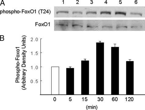
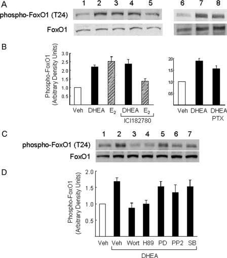
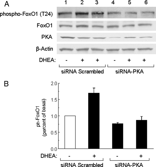
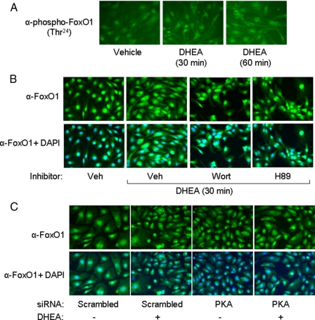
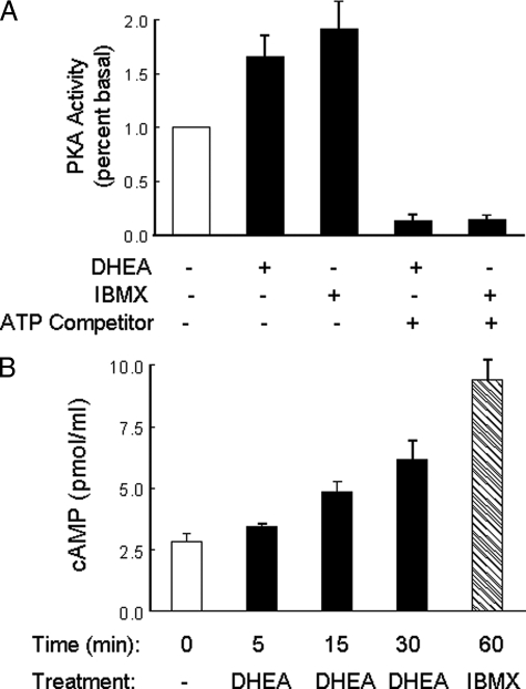
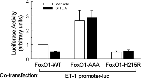
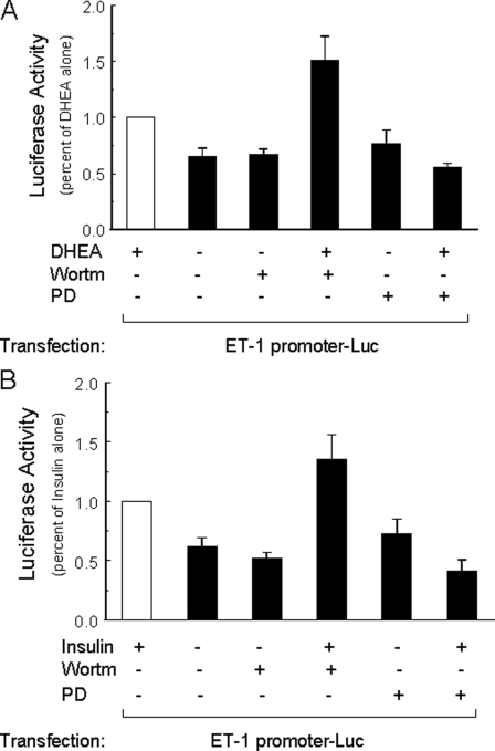
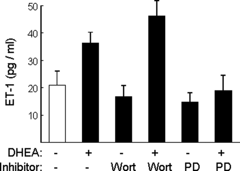
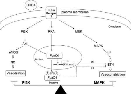
Dehydroepiandrosterone (DHEA) is an endogenous adrenal steroid hormone with controversial actions in humans. We previously reported that DHEA has opposing actions in endothelial cells to stimulate phosphatidylinositol (PI) 3-kinase/Akt/endothelial nitric-oxide synthase leading to increased production of nitric oxide while simultaneously stimulating MAPK-dependent secretion of the vasoconstrictor ET-1. In the present study we hypothesized that DHEA may stimulate PI 3-kinase-dependent phosphorylation of FoxO1 in endothelial cells to help regulate endothelial function. In bovine or human aortic endothelial cells (BAEC and HAEC), treatment with DHEA (100 nM) acutely enhanced phosphorylation of FoxO1. DHEA-stimulated phosphorylation of FoxO1 was inhibited by pretreatment of cells with wortmannin (PI 3-kinase inhibitor) or H89 (protein kinase A (PKA) inhibitor) but not ICI182780 (estrogen receptor blocker), or PD98059 (MEK (MAPK/extracellular signal-regulated kinase kinase) inhibitor). Small interfering RNA knockdown of PKA inhibited DHEA-stimulated phosphorylation of FoxO1. DHEA promoted nuclear exclusion of FoxO1 that was blocked by pretreatment of cells with wortmannin, H89, or by small interfering RNA knockdown of PKA. DHEA treatment of endothelial cells increased PKA activity and intracellular cAMP concentrations. Transfection of BAEC with a constitutively nuclear FoxO1 mutant transactivated a co-transfected ET-1 promoter luciferase reporter. Treatment of BAEC with DHEA inhibited transactivation of the ET-1 promoter reporter in cells overexpressing FoxO1. ET-1 promoter activity and secretion in response to DHEA treatment was augmented by PI 3-kinase blockade and inhibited by MAPK blockade. We conclude that DHEA stimulates phosphorylation of FoxO1 via PI 3-kinase- and PKA-dependent pathways in endothelial cells that negatively regulates ET-1 promoter activity and secretion. Balance between PI 3-kinase-dependent inhibition and MAPK-dependent stimulation of ET-1 secretion in response to DHEA may determine whether DHEA supplementation improves or worsens cardiovascular and metabolic function.


Figures

FIGURE 1.
FIGURE 1.
DHEA acutely stimulated phosphorylation of FoxO1 in endothelial cells in a time-dependent manner. A, BAECs were serum-starved overnight and then treated with DHEA (100 nm) for the durations indicated in panel B. Whole cell lysates were immunoblotted with antibodies against phospho-FoxO1 or FoxO1. Representative immunoblots are shown for experiments that were repeated independently four times. B, immunoblots from four independent experiments were quantified by scanning densitometry, and the amounts of phospho-FoxO1 were normalized to total FoxO1 in each lane. Results in the bar graph are the mean ± S.E. The amount of phospho-FoxO1 in cell lysates was significantly increased over basal after treatment of cells with DHEA for 30 (p < 0.0001) or 60 min (p < 0.04).
FIGURE 2.
FIGURE 2.
Phosphorylation of FoxO1 in response to DHEA in endothelial cells was not mediated through estrogen receptor or Gi but requires activation of PI 3-kinase and PKA. A, BAECs were serum-starved overnight and then treated with vehicle (lane 1), DHEA (100 nm, 30 min; lanes 2, 4, 7, and 8), or β-estradiol (E2, 20 nm, 5 min; lanes 3 and 5). Some cells were pretreated with estrogen receptor antagonist ICI182780 (10 μm, 30 min; lanes 4 and 5) or PTX (100 ng/ml, 2 h; lane 8) before DHEA or E2 treatment. Whole cell lysates were immunoblotted with antibodies against phospho-FoxO or FoxO1. Representative immunoblots are shown for experiments that were repeated independently three times. B, immunoblots from three independent experiments were quantified by scanning densitometry, and the amounts of phospho-FoxO1 were normalized to total FoxO1 in each lane. Results are plotted as a bar graph (mean ± S.E.). The amount of phospho-FoxO1 in the cell lysates was significantly increased over basal after treatment of cells with DHEA or β-estradiol (p < 0.001 and 0.005, respectively). Pretreatment with ICI182780 or PTX did not significantly block this effect of DHEA (p > 0.6 and 0.5, respectively). PTX, pertussis toxin. C, BAECs were serum-starved overnight and then treated with vehicle (lane 1) or DHEA (100 nm, 30 min; lanes 2–7). Some cells were pretreated for 1 h with the PI 3-kinase inhibitor wortmannin (100 nm; lane 3), PKA inhibitor H89 (25 μm; lane 4), MEK inhibitor PD98059 (25 μm; lane 5), Src-family kinase inhibitor PP2 (1 μm; lane 6), or p38 MAPK inhibitor SB203580 (10 μm; lane 7) before DHEA treatment. D, immunoblots from four independent experiments were quantified by scanning densitometry, and the amounts of phospho-FoxO1 were normalized to total FoxO1 in each lane. Results in the bar graphs are the mean ± S.E. The amount of phospho-FoxO1 in cell lysates was significantly increased over basal after treatment of cells with DHEA. Pretreatment with wortmannin or H89 (but not other inhibitors) significantly blocked this effect of DHEA (p < 0.02 when compared with DHEA treatment alone). Veh, vehicle.
FIGURE 3.
FIGURE 3.
siRNA knockdown of PKA inhibited DHEA-stimulated phosphorylation of FoxO1. A, HAECs were transfected with scrambled control siRNA (lanes 1–3) or siRNA specifically targeting PKA (lanes 4–6). 48 h after transfection, cells were serum-starved for 6 h and treated with vehicle or DHEA (100 nm, 30 min) as indicated. Whole cell lysates were immunoblotted with antibodies against phospho-FoxO1, FoxO1, PKA, or β-actin. Representative immunoblots are shown for experiments that were repeated independently three times. B, immunoblots from three independent experiments were quantified by scanning densitometry, and the amounts of phospho-FoxO1 were normalized to total FoxO1. Results in the bar graph are the mean ± S.E.
FIGURE 4.
FIGURE 4.
DHEA stimulated phosphorylation of FoxO1 in intact endothelial cells and promoted nuclear exclusion of FoxO1 in a PI 3-kinase- and PKA-dependent manner. A, serum-starved BAECs were treated with vehicle or DHEA (100 nm) for 30 or 60 min. Cells were immunostained with anti-phospho-FoxO1 antibody as described under “Materials and Methods.” B, serum-starved HAECs were treated with vehicle or DHEA (100 nm, 30 min) as indicated. Some cells were pretreated for 1 h with vehicle, wortmannin (100 nm), or H89 (25 μm) before DHEA treatment. Cells were immunostained with FoxO1 antibody (top panels). Co-staining of the same cells with FoxO1 antibody and 4,6-diamidino-2-phenylindole (DAPI) is shown in the bottom panels. Veh, vehicle. C, HAECs were transfected with siRNA targeting PKA or scrambled control siRNA. Two days after transfection, cells were serum-starved for 6 h and treated with vehicle or DHEA (100 nm, 30 min) as indicated. Cells were immunostained with FoxO1 antibody (top panels). Co-staining of the same cells with 4,6-diamidino-2-phenylindole is shown in the bottom panels.
FIGURE 5.
FIGURE 5.
DHEA increased PKA activity and intracellular cAMP in endothelial cells. A, serum-starved BAECs were treated for 1 h with vehicle, DHEA (100 nm), or IBMX (100 μm). Cytosolic fractions were isolated and assayed for PKA activity using a nonradioactive kinase activity assay kit as described under “Materials and Methods.” Results of PKA activity are presented as percent basal (mean ± S.E. of five independent experiments performed in duplicate). In some assays an ATP binding competitor (bisindolylmaleimide 1, 150 μm) was preincubated with cytosolic fractions for 1 min at room temperature. Both IBMX and DHEA significantly increased PKA activity when compared with basal levels (IBMX versus basal, p < 0.005; DHEA versus basal, p < 0.01). These effects of DHEA and IBMX were completely blocked by bisindolylmaleimide 1. B, serum-starved BAECs were treated with DHEA (100 nm for 0, 5, 15, or 30 min) or IBMX (100 μm, 1 h). cAMP concentrations were measured in cell lysates using an ELISA-based kit as described under “Materials and Methods.” DHEA treatment significantly increased intracellular cAMP concentrations at 30 min when compared with 0 min (p < 0.01). Data shown are the mean ± S.E. from seven independent experiments performed in duplicate.
FIGURE 6.
FIGURE 6.
DHEA inhibited the effect of FoxO1 to transactivate the ET-1 promoter. BAECs cultured in 24-well plates were co-transfected with an ET-1 promoter luciferase reporter construct and expression vectors for FoxO1-WT, FoxO1-AAA (constitutively nuclear mutant missing three Akt phosphorylation sites), or FoxO1-H215R (point mutant disrupting DNA binding site). One day after transfection, cells were serum-starved overnight and then treated with vehicle (open bars) or DHEA (100 nm, 8 h, closed bars) as indicated. A dual-luciferase reporter assay system was used to measure luciferase activity (mean ± S.E. of five independent experiments performed in triplicate), and data were normalized to the vehicle-treated FoxO1-WT group. DHEA treatment significantly blunted the effect of FoxO1-WT to transactivate the ET-1 promoter (p < 0.05).
FIGURE 7.
FIGURE 7.
Activation of ET-1 promoter in response to DHEA or insulin treatment was augmented by PI 3-kinase blockade and inhibited by MAPK blockade. BAECs grown in 24-well plates were transfected with ET-1 promoter luciferase reporter and renilla luciferase (internal control). One day later cells were serum-starved overnight and then treated for 8 h with vehicle, DHEA (100 nm), or insulin (100 nm) without or with wortmannin (100 nm), PD98059 (12.5 μm). Luciferase activity in each group was normalized to that in the group treated with DHEA alone (panel A, mean ± S.E. of eight independent experiments in triplicate) or insulin alone (panel B, mean ± S.E. of eight independent experiments in triplicate). When compared with cells treated with vehicle alone, ET-1 promoter activity was significantly increased in cells treated with either DHEA (panel A, p < 0.001) or insulin (panel B, p < 0.001). When compared with cells treated with DHEA or insulin alone, ET-1 promoter activity was further increased in cells co-treated with wortmannin (panel A, p < 0.03; panel B, p = 0.05) and inhibited in cells co-treated with PD98059 (panel A and B, p < 0.0001).
FIGURE 8.
FIGURE 8.
DHEA-stimulated increase in ET-1 secretion is augmented by PI 3-kinase blockade and inhibited by MAPK blockade. BAECs grown in 35-mm dishes were serum-starved overnight and then pretreated with vehicle, wortmannin (100 nm), or PD-98059 (12.5μm) for 1 h followed by treatment of DHEA (100 nm) or vehicle for 30 min. ET-1 concentration was measured in conditioned media using ELISA (mean ± S.E. of six independent experiments performed in duplicate). When compared with vehicle treatment, DHEA treatment significantly increased ET-1 secretion (p < 0.03), whereas wortmannin or PD98059 alone did not significantly change ET-1 concentration in conditioned media. When compared with DHEA treatment alone, wortmannin pretreatment enhanced the effect of DHEA to increase ET-1 secretion (p = 0.05), whereas PD98059 blocked the ability of DHEA to stimulate ET-1 secretion (p < 0.02).
FIGURE 9.
FIGURE 9.
Schematic model of signaling pathways that acutely regulate vascular actions of DHEA. As described previously (19), DHEA acutely activates PI 3-kinase/Akt/endothelial nitric-oxide synthase (eNOS) to stimulate production of the vasodilator NO. In addition, DHEA activates MAPK signaling to stimulate secretion of the vasoconstrictor ET-1. In the present study we demonstrated that DHEA acutely stimulates phosphorylation of FoxO1 using signaling pathways involving PKA and PI 3-kinase. Phosphorylated FoxO1 is excluded from the nucleus and translocated to the cytosol where it is unable to bind and activate the ET-1 promoter. Thus, a complex signaling network regulates opposing vascular actions of DHEA, and the net vasoactive action of DHEA is determined in part by the balance between PI 3-kinase- and MAPK-dependent signaling.

Similar articles

Cited by

References

    1. Lasley, B. L., Santoro, N., Randolf, J. F., Gold, E. B., Crawford, S., Weiss, G., McConnell, D. S., and Sowers, M. F. (2002) J. Clin. Endocrinol. Metab. 87 3760-3767 - PubMed
    1. Belanger, A., Candas, B., Dupont, A., Cusan, L., Diamond, P., Gomez, J. L., and Labrie, F. (1994) J. Clin. Endocrinol. Metab. 79 1086-1090 - PubMed
    1. Slowinska-Srzednicka, J., Zgliczynski, S., Soszynski, P., Makowska, A., Zgliczynski, W., Srzednicki, M., Bednarska, M., Chotkowska, E., Woroszylska, M., Ruzyllo, W., and Sadowski, Z. (1991) J. Intern. Med. 230 551-553 - PubMed
    1. Barrett-Connor, E., and Goodman-Gruen, D. (1995) Ann. N. Y. Acad. Sci. 774 259-270 - PubMed
    1. Johannes, C. B., Stellato, R. K., Feldman, H. A., Longcope, C., and McKinlay, J. B. (1999) J. Clin. Epidemiol. 52 95-103 - PubMed

Publication types