Review
. 2008 Oct;36(17):5482-515.
doi: 10.1093/nar/gkn517. Epub 2008 Aug 21.

Stability and kinetics of G-quadruplex structures

Affiliations
Review

Stability and kinetics of G-quadruplex structures

Andrew N Lane et al. Nucleic Acids Res. 2008 Oct.

Abstract

In this review, we give an overview of recent literature on the structure and stability of unimolecular G-rich quadruplex structures that are relevant to drug design and for in vivo function. The unifying theme in this review is energetics. The thermodynamic stability of quadruplexes has not been studied in the same detail as DNA and RNA duplexes, and there are important differences in the balance of forces between these classes of folded oligonucleotides. We provide an overview of the principles of stability and where available the experimental data that report on these principles. Significant gaps in the literature have been identified, that should be filled by a systematic study of well-defined quadruplexes not only to provide the basic understanding of stability both for design purposes, but also as it relates to in vivo occurrence of quadruplexes. Techniques that are commonly applied to the determination of the structure, stability and folding are discussed in terms of information content and limitations. Quadruplex structures fold and unfold comparatively slowly, and DNA unwinding events associated with transcription and replication may be operating far from equilibrium. The kinetics of formation and resolution of quadruplexes, and methodologies are discussed in the context of stability and their possible biological occurrence.


Figures
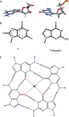
Figure 1.
Figure 1.
Chemical structures of G-quartets and quadruplexes. (A) Anticonformation (top left) and syn conformation (top right) of guanosine. (B) Inosine (left) and 7-deazaG (right) variations. (C) G-quartet with metal ion coordination to GO6.
Figure 2.
Figure 2.
Stacked quartets with coordinated monovalent ion. (A) Parallel stacked quartets with Na+ stabilization (purple spheres) from (d(TGGGGT)4), (B) parallel stacked quartets with K+ stabilization (green spheres) from (dA(GGGTTA)3GGG), (C) d(TGGGGT)4 stacking in space filling representation, (D) dA(GGGTTA)3GGG stacking in space filling representation. Loops have been removed from C and D for clarity.
Figure 3.
Figure 3.
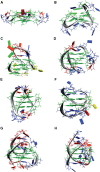
Observed quadruplex topologies. (A) All ‘parallel’ double chain reversal loops (dA(GGGTTA)3GGG: K+ form) (35), (B) all lateral loops d(GGTTGGTGTGGTTGG) (36), (C) lateral, lateral, double chain reversal loops d(GGGCGCGGGAGGAATTGGGCGGG) (37), (D) double chain reversal, lateral, lateral loops d(TTA(GGGTTA)3GGGA) (38), (E) lateral, diagonal, lateral loops dA(GGGTTA)3GGG (39), (F) diagonal, double chain reversal, diagonal loops (dGGTTTTGGCAGGGTTTTGGT) (40), (G) NMR-derived hybrid 1 (dAAA(GGGTTA)3GGGAA) (41) and (H) the NMR-derived hybrid 2 (dTTA(GGGTTA)3GGGTT) (42). Guanines are shown as green, thymine as blue and adenine as red oblongs.
Figure 4.
Figure 4.
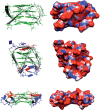
Topologies give rise to radically different structural appearance. Structures and electrostatic potential colored surfaces of the parallel (top), ‘basket’ lateral, diagonal, lateral loop (middle) and the all double chain reversal (bottom) topologies. The electrostatic surfaces are colored red (−10 kT/e) to blue (10 kT/e) and the bases are guanine in green, thymine in blue, and adenine in red.
Figure 5.
Figure 5.
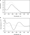
Absorbance and CD spectra. UV absorbance (A) and circular dichroic (B) spectra of the human telomere quadruplex sequence 5′AGGG(TTAGGG)3 in phosphate buffer (pH 7.0) containing 200 mM NaCl. Spectra obtained at 20°C are indicated by the solid line and correspond to the fully folded quadruplex form. Spectra obtained at 95°C are indicated by the dotted line, and correspond to the denatured, unfolded form.
Figure 6.
Figure 6.
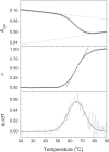
Thermal unfolding curves for the human intramolecular quadruplex. Transition curves for the denaturation of the Na+ form of the human telomere quadruplex sequence 5′AGGG(TTAGGG)3 in phosphate buffer (pH 7.0) containing 200 mM NaCl. (A) Absorbance at 295 nm versus temperature. The lines were calculated to fit the pre- and post-transition baselines. (B) Fraction of unfolded molecules (α) versus temperature after correction of the data in panel (A) for the sloping baselines and normalization. The straight line indicates the slope at the transition midpoint. (C) First derivative of the data in panel (B).
Figure 7.
Figure 7.
Whole-spectra melting data and the test of the two-state assumption. Thermal denaturation of the human telomere quadruplex sequence 5′AGGG(TTAGGG)3 in a solution containing 0.185 M NaCl is shown as monitored by UV absorbance (A) or CD (B). The corresponding two-wavelength parametric plots to test the two-state assumption (144) are shown in (C and D). The nonlearity of the the data in panels C and D indicate that the denaturation of the quadruplex is not a simple two-state process, and the intermediate states must be included in the reaction mechanism.
Figure 8.
Figure 8.
Thermal profiles for two folding pathways. The populations of states in two possible pathways as described in the text was calculated. Model (i) Two species connected by unfolded state: N1⇔D + N2 ⇔ D. The reference temperature, Tref = 273 K. The equilibrium constant K0 for unfolding at 273 K = 1E − 5. The unfolding enthalpies were Δ H1 (N1) = 40 kcal mol−1, Δ H2(N2) = 60 kcal mol−1. For these parameters, Tm1 = 324 K Tm2 = 305 K. (A) Populations as a function of temperature. Red square: state D; open black circles: state N2; filled blue circles: state N1. (B) Changes in absorbance as a function of temperature for εN1 = εN2 (filled red squares), εN1 = 1.2, εN2 = 1 (open blue squares) and εN1 = 1, εN2 = 1.2 (open black circles). Best fit to a single transition with εN1 = εN2: ΔH = 40 kcal mol−1; K0 = 9.7 E−6 at 273 K; Tm = 324 K. Model (ii) sequential unfolding NK1 IK2 K1 = K2 = 1E−5 at 273 K, Δ H1 = 30 kcal mol−1, Δ H2 = 20 kcal mol−1. For these parameters, Tm1 = 305 K, Tm2 = 324 K. (C) Populations of N (red squares), I (black circles) and U (blue squares). The populations of N and I are equal at 305 K, and N is dominant at low temperature. (D) Thermal melting profile using the populations in C, and difference absorption coefficients of (a) ΔεN = ΔεI = 1 (blue squares) (b) ΔεN = 1.2 ΔεI = 1 (black circles); (c) ΔεN = 1 ΔεI = 1.2 (blue squares). The fits for conditions a and b to a single folding transition are shown as thin continuous lines, and the recovered parameter values were: (A) K(273) = 8.7 E−6, ΔH = 40.5 ± 0.07 kcal mol−1, Δε = 1.0, R2 = 0.99997. (B) K(273) = 1.68 E−4; ΔH = 31.6 ± 0.3 kcal mol−1, Δε = 1.19, R2 = 0.99895. Parameter estimates are thus unreliable if the wrong model is used, even where the data appear as a simple transition.
Figure 9.
Figure 9.
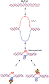
Formal model for formation of an internal G-quadruplex or other intramolecular structure. Duplex DNA with a G-quadruplex potential sequence (top) unwinds and histones redistribute, leading to base-pair dissociation and formation of an open loop (middle). The G-rich strand forms a unimolecular G-quadruplex structure in the presence of a single-stranded complementary C-rich strand. This potentially could form an intramolecular i-motif as shown (third from top). The quadruplex and/or the C-rich strand may be stabilized by proteins (as shown at bottom) or other ligands. The binding energy required to overcome the unwinding is discussed in the text.

References

    1. Huppert JL, Balasubramanian S. Prevalence of quadruplexes in the human genome. Nucleic Acids Res. 2005;33:2908–2916. - PMC - PubMed
    1. Huppert JL, Balasubramanian S. G-quadruplexes in promoters throughout the human genome. Nucleic Acids Res. 2007;35:406–413. - PMC - PubMed
    1. Cech TR. Beginning to understand the end of the chromosome. Cell. 2004;116:273–279. - PubMed
    1. Chan S, Blackburn EH. Telomeres and telomerase. Philos. Trans. R. Soc. Lond. B-Biol. Sci. 2004;359:109–121. - PMC - PubMed
    1. Blackburn EH. Switching and signaling at the telomere. Cell. 2001;106:661–673. - PubMed

Publication types