. 2009 Jan;150(1):519-29.
doi: 10.1210/en.2008-0878. Epub 2008 Aug 21.

Characterization of thyrotropin receptor antibody-induced signaling cascades

Affiliations

Characterization of thyrotropin receptor antibody-induced signaling cascades

Syed A Morshed et al. Endocrinology. 2009 Jan.

Abstract

The TSH receptor (TSHR) is constitutively active and is further enhanced by TSH ligand binding or by stimulating TSHR antibodies (TSHR-Abs) as seen in Graves' disease. TSH is known to activate the thyroid epithelial cell via both Galphas-cAMP/protein kinase A/ERK and Galphaq-Akt/protein kinase C coupled signaling networks. The recent development of monoclonal antibodies to the TSHR has enabled us to investigate the hypothesis that different TSHR-Abs may have unique signaling imprints that differ from TSH ligand itself. We have, therefore, performed sequential studies, using rat thyrocytes (FRTL-5, passages 5-20) as targets, to examine the signaling pathways activated by a series of monoclonal TSHR-Abs in comparison with TSH itself. Activation of key signaling molecules was estimated by specific immunoblots and/or enzyme immunoassays. Continuing constitutive TSHR activity in thyroid cells, deprived of TSH and serum for 48 h, was demonstrated by pathway-specific chemical inhibition. Under our experimental conditions, TSH ligand and TSHR-stimulating antibodies activated both Galphas and Galphaq effectors. Importantly, some TSHR-blocking and TSHR-neutral antibodies were also able to generate signals, influencing primarily the Galphaq effectors and induced cell proliferation. Most strikingly, antibodies that used the Galphaq cascades used c-Raf-ERK-p90RSK as a unique signaling cascade not activated by TSH. Our study demonstrated that individual TSHR-Abs had unique molecular signatures which resulted in sequential preferences. Because downstream thyroid cell signaling by the TSHR is both ligand dependent and independent, this may explain why TSHR-Abs are able to have variable influences on thyroid cell biology.


Figures
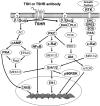
Figure 1
Figure 1
Simplified diagrammatic illustration of the major signaling pathways evaluated in the current studies. Note the five pathways from left to right: cAMP/PKA/ERK or PKA/CREB, PI3/Akt/mTOR, PKC/NFκB, PKC/c-raf/ERK/p90RSK, and Ras/c-Raf/ERK. In practice, many of these pathways interact, and the diagram is for illustrative purposes only.
Figure 2
Figure 2
Intracellular cAMP generation in quiescent FRTL-5 cells. Increasing concentrations of TSH (0.1, 1.0, and 10 mU/ml) or TSHR-stimulating monoclonal antibodies (0.1, 1.0, and 10 μg/ml) induced cAMP during a 60-min exposure as illustrated by fold increases in picomoles per milliliter (A). At fixed concentrations of TSH (1 mU/ml) and antibodies (1 μg/ml), an hour of stimulation produced significantly increased levels of cAMP (TSH, P < 0.007; M22, P < 0.01; R12, P < 0.01, MS1, P < 0.001; B2, P < 0.009; T8, P < 0.035) when compared with the medium alone or a control antibody. B, Detailed changes of selected stimulators or blocker over time. C, cAMP responses in JPO9 cells produced by stimulators or blocker as in B. B2 dose-response is also shown in C. These demonstrate endogenous (basal) cAMP (without stimulation) and stimulated cAMP [using 1 mU/ml TSH or 1 μg/ml monoclonal antibody (mAb)]. The individual antibodies are labeled according to Table 1 (R12, RSR-12; B2, RSR-B2; T8, Tab-8; 7G, 7G10; T16, Tab-16).
Figure 3
Figure 3
Induction of FRTL-5 cell proliferation. Quiescent and starved cells were stimulated with increasing concentrations of TSH (0.1, 1.0, and 10 mU/ml) or TSHR antibodies (0.1, 1.0, and 10 μg/ml) in the basal medium. At a fixed concentration, TSH (1 mU/ml) stimulating and blocking antibodies (1 μg/ml) demonstrated significantly higher cell proliferation (TSH, P < 0.0004; M22, P < 0.0005; R12, P < 0.0004; MS1, P < 0.003; B2, P < 0.0008; T8; P < 0.001) than medium alone (basal) or a control antibody (Cont; 1 μg/ml). One of the neutral (T16; 1 μg/ml) antibodies also showed significantly higher proliferation (P < 0.001), whereas 7G showed suppressive effects (P < 0.02) at a higher concentration (10 μg/ml). These experiments were repeated twice in triplicate.
Figure 4
Figure 4
The constitutive activity of signaling molecules in untreated FRTL-5 cells. Starved quiescent cells were left untreated for multiple different time points, and their signaling activities were examined by immunoblotting with phosphospecific antibodies. Signaling molecule activity decreased in a time-dependent manner over 60 min as demonstrated by repeated experiments. A, Representative immunoblots containing phospho- or non-phosphoproteins. The lowercase letter p in front of each molecule indicates phosphoprotein detection. B, Densitometric quantitation of each band on the autoradiographs produced from immunoblots. Quiescent FRTL-5 cells were treated with a fixed concentration of specific kinase inhibitors as described in Materials and Methods. Both reductions and increases in phosphorylated proteins were observed by these specific inhibitors. Of note, unlike suppressing Akt, PI3-kinase inhibitor (Ly294002; 1 mm) activated PKC, CREB, and ERK/p90RSK (C and D).
Figure 5
Figure 5
TSH uses both Gαs and Gαq pathways. In 60 min, TSH repeatedly activated Akt, PKC, and CREB but not PKA and ERK as illustrated in these representative immunoblots (A). Densitometric quantitation of each band was plotted as fold activation (B), which indicated that the activity of the phosphoproteins had either reduced or increased.
Figure 6
Figure 6
TSHR-stimulating antibodies activated TSH signaling pathways. Increasing concentrations (0.1, 1.0, and 10 μg/ml) of TSHR-stimulating antibodies MS1, R12, and M22 over a 60-min incubation showed similar changes in signaling molecules as seen with TSH ligand in these representative immunoblots. M22 produced dose-dependent significant increases in Akt, PKA, and CREB activities compared with TSH and the other antibodies. These dose-response studies were repeated twice.
Figure 7
Figure 7
PKA and PKC activity assessed by EIA. In our model system, TSH (1 mU/ml) did not activate PKA enzyme activity, whereas the M22 TSHR-stimulating antibody (1 μg/ml) did demonstrate a highly significant increase (P < 0.0001). R12 (P < 0.013) and MS1 (P < 0.03) stimulating antibodies showed significantly reduced activities. Both neutral antibodies also exhibited similar effects (T16 and 7G, P < 0.02). Although blocking antibodies did not produce any significant effects. In contrast, both TSH (P < 0.0005) and TSHR-stimulating antibodies increased PKC (M22, P < 0.001; R12, P < 0.003) activity significantly. One of blocking (B2, P < 0.0001) and both neutral (T16, P < 0.0001; 7G, P < 0.0002) antibodies also demonstrated a significant increase in PKC activity when compared with the medium alone (basal) or a control monoclonal antibody (cont). The experiments were repeated three times and confirmed the illustrated findings.
Figure 8
Figure 8
Increasing concentrations (0.1, 1.0, and 10 μg/ml) of TSHR blocking (A) and neutral (B) antibodies over a 60-min incubation showed variable changes in signaling molecules as shown in these representative immunoblots. The data (representing one set of experiments repeated three times) showed different signaling cascades induced by blocking and neutral antibodies. For example, the B2 blocking antibody increased several signaling molecules, the most important of which appeared to be the c-Raf/ERK pathway, whereas the 7G neutral TSHR antibody suppressed signaling molecules such as c-Raf/ERK. T16, also a neutral antibody, showed increasing activities of many signaling molecules including Akt, PKC, CREB, and c-Raf/ERK/p90RSK. By contrast, an isotype control monoclonal antibody (IgG2) did not produce such dose-dependent activities.

References

    1. Kimura T, Van KA, Golstein J, Fusco A, Dumont JE, Roger PP 2001 Regulation of thyroid cell proliferation by TSH and other factors: a critical evaluation of in vitro models. Endocr Rev 22:631–656 - PubMed
    1. Medina DL, Santisteban P 2000 Thyrotropin-dependent proliferation of in vitro rat thyroid cell systems. Eur J Endocrinol 143:161–178 - PubMed
    1. Bachrach LK, Eggo MC, Mak WW, Burrow GN 1985 Phorbol esters stimulate growth and inhibit differentiation in cultured thyroid cells. Endocrinology 116:1603–1609 - PubMed
    1. Roger PP, Reuse S, Servais P, Van HB, Dumont JE 1986 Stimulation of cell proliferation and inhibition of differentiation expression by tumor-promoting phorbol esters in dog thyroid cells in primary culture. Cancer Res 46:898–906 - PubMed
    1. Morrison DK, Cutler RE 1997 The complexity of Raf-1 regulation. Curr Opin Cell Biol 9:174–179 - PubMed

Publication types