

Preoperative grading of presumptive low-grade astrocytomas on MR imaging: diagnostic value of minimum apparent diffusion coefficient
- PMID: 18719036
- PMCID: PMC8118927
- DOI: 10.3174/ajnr.A1254
Preoperative grading of presumptive low-grade astrocytomas on MR imaging: diagnostic value of minimum apparent diffusion coefficient
Abstract
Background and purpose: Histopathologic grade of glial tumors is inversely correlated with the minimum apparent diffusion coefficient (ADC). We assessed the diagnostic values of minimum ADC for preoperative grading of supratentorial astrocytomas that were diagnosed as low-grade astrocytomas on conventional MR imaging.
Materials and methods: Among 118 patients with astrocytomas (WHO grades II-IV), 16 who showed typical MR imaging findings of low-grade supratentorial astrocytomas on conventional MR imaging were included. All 16 patients underwent preoperative MR imaging and diffusion-weighted imaging. The minimum ADC value of each tumor was determined from several regions of interest in the tumor on ADC maps. To assess the relationship between the minimum ADC and tumor grade, we performed the Mann-Whitney U test. A receiver operating characteristic (ROC) analysis was used to determine the cutoff value of the minimum ADC that had the best combination of sensitivity and specificity for distinguishing low- and high-grade astrocytomas.
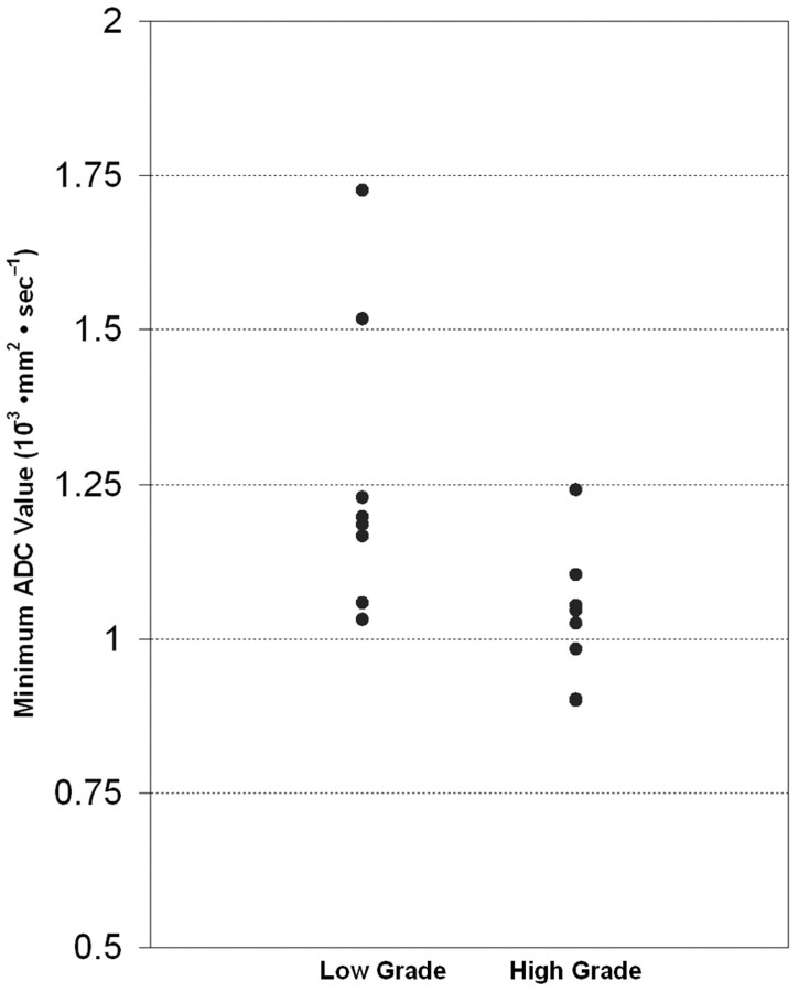
Results: Eight of the 16 patients (50%) were confirmed as having high-grade astrocytomas (WHO grades III and IV), and the other 8 patients were confirmed as having low-grade astrocytomas (WHO grade II). The median minimum ADC of the high-grade astrocytoma (1.035 x 10(-3) mm(2) . sec(-1)) group was significantly lower than that of the low-grade astrocytoma group (1.19 x 10(-3) mm(2) . sec(-1)) (P = .021). According to the ROC analysis, the cutoff value of 1.055 x 10(-3) mm(2) . sec(-1) for the minimum ADC generated the best combination of sensitivity (87.5%) and specificity (79%) (P = .021).
Conclusion: Measuring minimum ADC can provide valuable diagnostic information for the preoperative grading of presumptive low-grade supratentorial astrocytomas.
Figures
References
-
- Bulakbasi N, Guvenc I, Onguru O, et al. The added value of the apparent diffusion coefficient calculation to magnetic resonance imaging in the differentiation and grading of malignant brain tumors. J Comput Assist Tomogr 2004;28:735–46 - PubMed
-
- Yang D, Korogi Y, Sugahara T, et al. Cerebral gliomas: prospective comparison of multivoxel 2D chemical-shift proton MR spectroscopy, echoplanar perfusion and diffusion-weighted MRI. Neuroradiology 2002;44:656–66 - PubMed
-
- Higano S, Yun X, Kumabe T, et al. Malignant astrocytic tumors: clinical importance of apparent diffusion coefficient in prediction of grade and prognosis. Radiology 2006;241:839–46 - PubMed
-
- Tervonen O, Forbes G, Scheithauer BW, et al. Diffuse fibrillary astrocytomas: correlation of MRI features with histopathologic parameters and tumor grade. Neuroradiology 1992;34:173–78 - PubMed
MeSH terms
LinkOut - more resources
Full Text Sources
Medical