. 2008 Aug 26;105(34):12429-34.
doi: 10.1073/pnas.0806585105. Epub 2008 Aug 21.

Beyond tumor necrosis factor receptor: TRADD signaling in toll-like receptors

Affiliations

Beyond tumor necrosis factor receptor: TRADD signaling in toll-like receptors

Nien-Jung Chen et al. Proc Natl Acad Sci U S A. .

Abstract

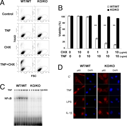
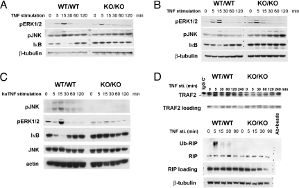
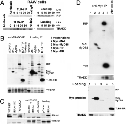
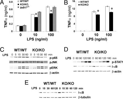
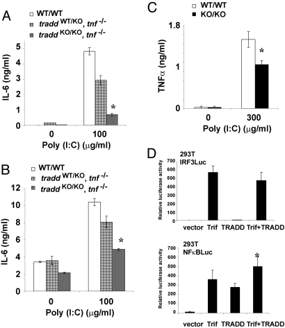
Tumor necrosis factor receptor 1-associated death domain protein (TRADD) is the core adaptor recruited to TNF receptor 1 (TNFR1) upon TNFalpha stimulation. In cells from TRADD-deficient mice, TNFalpha-mediated apoptosis and TNFalpha-stimulated NF-kappaB, JNK, and ERK activation are defective. TRADD is also important for germinal center formation, DR3-mediated costimulation of T cells, and TNFalpha-mediated inflammatory responses in vivo. TRADD deficiency does not enhance IFNgamma-induced signaling. Importantly, TRADD has a novel role in TLR3 and TLR4 signaling. TRADD participates in the TLR4 complex formed upon LPS stimulation, and TRADD-deficient macrophages show impaired cytokine production in response to TLR ligands in vitro. Thus, TRADD is a multifunctional protein crucial both for TNFR1 signaling and other signaling pathways relevant to immune responses.


Conflict of interest statement

The authors declare no conflict of interest.

Figures

Fig. 1.
Fig. 1.
TRADD is indispensable for TNFα-induced cell death and TNFα-mediated NF-κB activation. Primary MEFs from traddWT/WT and traddKO/KO mice were treated for 24 h with TNFα alone (10 ng/ml), CHX alone (1–10 μg/ml), or TNFα plus CHX. Viability was assessed by 7-AAD staining and flow cytometry. (A) Representative flow cytometric data with 1 μg/ml CHX. (B) Bar graph summary of all flow cytometric data. Results shown are the mean viability ± SD of triplicate determinations. **, P < 0.01, Student's t test. For A and B, data are representative of at least three independent experiments. (C) Gel mobility shift assay. MEFs from traddWT/WT and traddKO/KO mice were stimulated with 10 ng/ml TNFα for the indicated times, and NF-κB activation was evaluated by EMSA. (D) RelA/p65 translocation. The MEFs in A were subjected to immunofluorescent staining to detect RelA/p65 translocation into the nucleus. MEFs were also treated with LPS (500 ng/ml) or IL-1β (10 ng/ml), and RelA/p65 translocation was assessed.
Fig. 2.
Fig. 2.
Alterations to TNFR1 signaling and complex formation in the absence of TRADD. (A–C) ERK, JNK, and IκB. traddWT/WT and traddKO/KO MEFs (A) and BM-Macs (B) were stimulated with 10 ng/ml murine TNFα (A and B) or 10 ng/ml human TNF (C) for the indicated times, and ERK and JNK phosphorylation and IκB degradation were assessed by Western blotting. β-tubulin and actin were used as loading controls. (D) TNFR1 complex composition. traddWT/WT and traddKO/KO MEFs were stimulated with 10 ng/ml TNFα for the indicated times, and the TNFR1 complex was isolated by IP. TRAF2, RIP, and ubiquitinated RIP (Ub-RIP) within the TNFR1 complex were detected by Western blotting. Data shown are representative of at least three independent experiments.
Fig. 3.
Fig. 3.
TRADD is recruited to the TLR4 signaling complex upon LPS stimulation. (A) TRADD participates in the TLR4 complex. RAW cells were stimulated with LPS (100 ng/ml) for the indicated times, and lysates were subjected to IP with anti-TLR4. TRADD and RIP were identified by Western blotting to be components of the TLR4 complex. (B and C) TRADD associates with TLR4-TIR but not IRAK4. 293T cells were cotransfected with a TRADD-expressing vector plus vectors expressing the indicated Myc-tagged mediators. Proteins associating with TRADD were identified by TRADD-IP and anti-Myc Western blotting. IRAK4KO was used as a negative control; pCDNA3 and pBABE were used as empty vector controls. (D) TLR4-TIR associates with TRADD; reciprocal IP involving anti-Myc IP and anti-TRADD Western blotting. For A–D, data are representative of at least two independent experiments.
Fig. 4.
Fig. 4.
TRADD is involved in TLR4 signaling and TLR4 mediated cytokine production. (A and B) Reduced LPS-stimulated TNFα production. traddWT/WT, traddKO/KO, and tnfr1−/− BM-Macs (A) and traddWT/WT and traddKO/KO peritoneal macrophages (B) were stimulated with LPS (10, 100, or 300 ng/ml) for 24 h, and TNFα in the culture supernatants was evaluated by ELISA. *, P < 0.05, Student's t test. (C–E) Reduced LPS-stimulated NF-κB and MAPK activation. traddWT/WT and traddKO/KO BM-Macs were stimulated with 100 ng/ml LPS for the indicated times. The phosphorylation of p65, ERK, JNK, and STAT1, and IκB degradation were assessed by Western blotting. β-actin and β-tubulin were used as loading controls. For A–E, data are representative of at least two independent experiments.
Fig. 5.
Fig. 5.
TRADD is involved in TLR3 mediated cytokine production. (A and B) Reduced poly(I:C)-stimulated IL6 production. traddWT/WT, traddWT/WTtnf−/− and traddKO/KOtnf−/− BM-Macs (A) and lung fibroblasts (B) were stimulated with 100 μg/ml poly(I:C), and IL-6 in the culture supernatants was evaluated 24 h later by ELISA. (C) Reduced poly(I:C)-stimulated TNFα production. traddWT/WT and traddKO/KO peritoneal macrophages were stimulated with 300 μg/ml poly(I:C), and TNFα in the culture supernatants was evaluated 24 h later by ELISA. (D) TRADD augments TRIF-dependent NF-κB activation. 293T cells were transfected with either an NF-κB or IRF3 luciferase reporter in the presence of TRADD and/or TRIF expression vectors. Results are expressed as luciferase activity relative to β-galactosidase activity ± SD. For A–D, *, P < 0.05, Student's t test. Data are representative of at least two independent experiments.

References

    1. Aggarwal BB. Signalling pathways of the TNF superfamily: A double-edged sword. Nat Rev Immunol. 2003;3:745–756. - PubMed
    1. Hsu H, Xiong J, Goeddel DV. The TNF receptor 1-associated protein TRADD signals cell death and NF-kappa B activation. Cell. 1995;81:495–504. - PubMed
    1. Chen G, Goeddel DV. TNF-R1 signaling: A beautiful pathway. Science. 2002;296:1634–1635. - PubMed
    1. Mak TW, Yeh WC. Signaling for survival and apoptosis in the immune system. Arthritis Res 4 Suppl. 2002;3:S243–252. - PMC - PubMed
    1. Chinnaiyan AM, et al. Signal transduction by DR3, a death domain-containing receptor related to TNFR-1 and CD95. Science. 1996;274:990–992. - PubMed

Publication types