. 2008 Aug 26;105(34):12176-81.
doi: 10.1073/pnas.0803626105. Epub 2008 Aug 21.

Structure of apolipoprotein A-I in spherical high density lipoproteins of different sizes

Affiliations

Structure of apolipoprotein A-I in spherical high density lipoproteins of different sizes

R A Gangani D Silva et al. Proc Natl Acad Sci U S A. .

Abstract

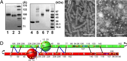
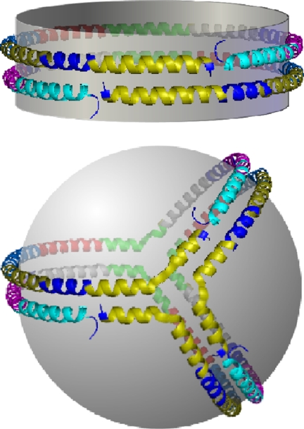
Spherical high density lipoproteins (HDL) predominate in human plasma. However, little information exists on the structure of the most common HDL protein, apolipoprotein (apo) A-I, in spheres vs. better studied discoidal forms. We produced spherical HDL by incubating reconstituted discoidal HDL with physiological plasma-remodeling enzymes and compared apoA-I structure in discs and spheres of comparable diameter (79-80 and 93-96 A). Using cross-linking chemistry and mass spectrometry, we determined that the general structural organization of apoA-I was overall similar between discs and spheres, regardless of diameter. This was the case despite the fact that the 93 A spheres contained three molecules of apoA-I per particle compared with only two in the discs. Thus, apoA-I adopts a consistent general structural framework in HDL particles-irrespective of shape, size and the number of apoA-Is present. Furthermore, a similar cross-linking pattern was demonstrated in HDL particles isolated from human serum. We propose the first experiment-based molecular model of apoA-I in spherical HDL particles. This model provides a new foundation for understanding how apoA-I structure modulates HDL function and metabolism.


Conflict of interest statement

The authors declare no conflict of interest.

Figures

Fig. 1.
Fig. 1.
Structural analysis of D79 and S80 particles. (A) Native PAGGE (8–25%) of D79 (lane 1) and S80 (lane 3) particles. An 8–25% SDS/PAGGE analysis of cross-linked S80 after separation into monomeric (lane 5) and dimeric (lane 6) forms by gel filtration chromatography is also shown. Cross-linking was carried out at a 1:10 molar ratio of apoA-I: BS3 at 1 mg/ml apoA-I concentration. All gels were stained with Coomassie blue. (B and C) Negative stain electron micrographs of D79 and S80 particles. (Scale bars: 50 nm.) (D) Experimental cross-links compatible with 5/5 molecular registry of the double-belt model are shown as solid lines. The apoA-I molecules have been drawn as if they have been peeled off the edge of an HDL disk and laid flat. Cross-links in black are common to both D79 and S80 particles; those in blue are found in D79 only and those in gray are found in S80 only. Putative apoA-I helical segments are numbered according to Roberts et al. (31). Locations of the 21 Lys residues in apoA-I are identified as orange dots. All experimental cross-links found in D79 and S80 are listed in Table S1.
Fig. 2.
Fig. 2.
Structural analysis of D96 and S93 particles. (A) Native PAGGE (8–25%) of D96 (lane 1) and S93 (lane 2). An 8–25% SDS/PAGGE analysis of cross-linked D96 (lane 6) and S93 (lane 7) is also shown. The particles were cross-linked at a 1:100 molar ratio of apoA-I: BS3 at 1 mg/ml apoA-I for PAGGE analysis to avoid partial cross-linking and to detect the highest MW bands. Lipid-free apoA-I (lane 4) and lipid-free apoA-I cross-linked under the same conditions (lane 5) is shown for comparison. All gels were stained with Coomassie Blue. (B and C) Negative stain EM images of D96 and S93 particles, respectively. (Scale bars: 50 nm.) (D) Experimental cross-links compatible with 5/5 molecular registry of the double-belt model are shown as for Fig. 1. Cross-linker color coding is the same as in Fig. 1.
Fig. 3.
Fig. 3.
Potential molecular arrangements of three apoA-I molecules on the larger HDL spherical particles. The classical 5/5 double belt model proposed for D96 is shown first with two molecules of apoA-I shown in red and green (model A). The small spheres represent the N-terminal 44 aa of each molecule. Model B shows two molecules of apoA-I arranged in a double belt as in the discs with a third molecule arranged as a hairpin on one hemisphere. Model C has all three molecules arranged in an antiparallel fashion at the particle equator, i.e., triple belt. Model D, called the trefoil model, was generated by splitting the right hand half of two molecules of apoA-I in the double belt 60° out of the plane of the disk. Then a third molecule, bent the same way, was inserted.
Fig. 4.
Fig. 4.
Molecular comparison of the apoA-I interactions in a disk double belt vs. the spherical trefoil. Each putative helical domain shown in Fig. 1 is represented as a separate color; helix 1, teal; helix 2 purple; helix 3, dark blue; helix 4, gray; helix 5, green; helix 6, red; helix 7, light blue; helix 8, dark yellow; helix 9, navy blue; helix 10, yellow. Notice that all helix to helix interactions present in the double belt between two molecules of apoA-I in the disk are also present between three apoA-I molecules in the trefoil. For example, helix 10 of all three molecules in the trefoil (the yellow one in front) can interact with helix 10 of the other molecules in the same way as the double belt. Another example is the similar arrangements of helix 9 (navy blue) and helix 1 (teal) in both models.

References

    1. Curtiss LK, Valenta DT, Hime NJ, Rye KA. What is so special about apolipoprotein AI in reverse cholesterol transport? Arterioscler Thromb Vasc Biol. 2006;26:12–19. - PubMed
    1. Brouillette CG, Anantharamaiah GM, Engler JA, Borhani DW. Structural models of human apolipoprotein A-I: A critical analysis and review. Biochim Biophys Acta. 2001;1531:4–46. - PubMed
    1. Segrest JP, et al. A detailed molecular belt model for apolipoprotein A-I in discoidal high density lipoprotein. J Biol Chem. 1999;274:31755–31758. - PubMed
    1. Jonas A. In: Biochemistry of Lipids, Lipoproteins and Membranes. Vance DE, Vance JE, editors. Vol. 483. Elsevier Science; 2002. p. 504.
    1. Borhani DW, Rogers DP, Engler JA, Brouillette CG. Crystal structure of truncated human apolipoprotein A-I suggests a lipid-bound conformation. Proc Natl Acad Sci USA. 1997;94:12291–12296. - PMC - PubMed

Publication types

LinkOut - more resources