. 2008 Oct;14(10):2149-58.
doi: 10.1261/rna.1094208. Epub 2008 Aug 21.

lsm1 mutations impairing the ability of the Lsm1p-7p-Pat1p complex to preferentially bind to oligoadenylated RNA affect mRNA decay in vivo

Affiliations

lsm1 mutations impairing the ability of the Lsm1p-7p-Pat1p complex to preferentially bind to oligoadenylated RNA affect mRNA decay in vivo

Ashis Chowdhury et al. RNA. 2008 Oct.

Abstract

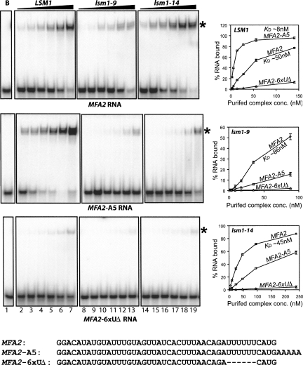
The poly(A) tail is a crucial determinant in the control of both mRNA translation and decay. Poly(A) tail length dictates the triggering of the degradation of the message body in the major 5' to 3' and 3' to 5' mRNA decay pathways of eukaryotes. In the 5' to 3' pathway oligoadenylated but not polyadenylated mRNAs are selectively decapped in vivo, allowing their subsequent degradation by 5' to 3' exonucleolysis. The conserved Lsm1p-7p-Pat1p complex is required for normal rates of decapping in vivo, and the purified complex exhibits strong binding preference for oligoadenylated RNAs over polyadenylated or unadenylated RNAs in vitro. In the present study, we show that two lsm1 mutants produce mutant complexes that fail to exhibit such higher affinity for oligoadenylated RNA in vitro. Interestingly, these mutant complexes are normal with regard to their integrity and retain the characteristic RNA binding properties of the wild-type complex, namely, binding near the 3'-end of the RNA, having higher affinity for unadenylated RNAs that carry U-tracts near the 3'-end over those that do not and exhibiting similar affinities for unadenylated and polyadenylated RNAs. Yet, these lsm1 mutants exhibit a strong mRNA decay defect in vivo. These results underscore the importance of Lsm1p-7p-Pat1p complex-mRNA interaction for mRNA decay in vivo and imply that the oligo(A) tail mediated enhancement of such interaction is crucial in that process.


Figures

FIGURE 1.
FIGURE 1.
Lsm1p-7p-Pat1p complexes isolated from lsm1-9 and lsm1-14 mutants show a band pattern similar to the wild-type complex in SDS-PAGE. Lsm1p-7p-Pat1p complexes purified from LSM1, lsm1-9, and lsm1-14 cells (lanes 1,2,3, respectively) as described in Materials and Methods were subjected to SDS-PAGE separation followed by visualization of the bands by silver staining. Positions of the molecular weight markers are shown on the left. Identity of the protein bands is indicated on the right wherein asterisks mark the higher mobility species of Pat1p (Chowdhury et al. 2007).
FIGURE 2.
FIGURE 2.
Mutant Lsm1p-7p-Pat1p complexes purified from lsm1-9 and lsm1-14 cells are unable to recognize oligoadenylated status of RNAs. Gel mobility shift assays were carried out as described in Materials and Methods using MFA2, MFA2-A5, or MFA2-6xUΔ (phosphorimages in the first, second, and third panels, respectively, from top) RNA with BSA (lane 1) or increasing concentrations of the complex purified from LSM1 (lanes 2–7), lsm1-9 (lanes 8–13), or lsm1-14 (lanes 14–19) cells. RNA binding was quantitated using a PhosphorImager, and plots of the fraction of RNA bound versus the concentration of the purified complex are shown on the right. Sequences of the RNAs used are shown in the bottom. Asterisks indicate the position of the gel shifted RNA in the gel pictures.
FIGURE 3.
FIGURE 3.
Inability of the mutant complexes to recognize the 3′-oligo(A) tail is not RNA specific. Gel mobility shift assays were carried out as described in Materials and Methods using PGK1 (lanes 1–7) or PGK1-A5 (lanes 8–14) RNA with BSA (lanes 1,14) or increasing concentrations of the complex purified from LSM1, lsm1-9, or lsm1-14 cells (phosphorimages in top, middle, and bottom panels, respectively). Plots of the fraction of RNA bound versus the concentration of the purified complex are shown on the right. Sequences of the RNAs used are shown in the bottom.
FIGURE 4.
FIGURE 4.
Binding of MFA2 RNA by the mutant complexes is also impaired by addition of a pentanucleotide non-oligo(A) tail at the 3′-end. Gel mobility shift assays were carried out as described in Materials and Methods using MFA2 or MFA2-CAGAC RNAs (indicated below the phosphorimages) with BSA or increasing concentrations of the complex purified from LSM1, lsm1-9, or lsm1-14 cells (indicated above the phosphorimages). Fraction of RNA bound in the different reactions (quantitated using a PhosphorImager) is shown as a bar diagram in the lower panel. The numbers of the lanes in the upper panel that correspond to each pair of bars in the bar diagram are shown below the bars. In each set of reactions (LSM1, lsm1-9, or lsm1-14), the fraction of RNA bound is normalized to the value obtained with the MFA2 RNA at the highest concentration of the purified complex used.
FIGURE 5.
FIGURE 5.
Like the wild-type complex, the mutant complexes also bind near the 3′-end of the RNA and exhibit similar affinities for unadenylated and polyadenylated RNAs. (A) Mutant complex purified from lsm1-14 cells binds near the 3′-end of the RNA. Radiolabeled MFA2(u) RNA was annealed to DNA oligonucleotides oST214, oST215, oST216, a nonspecific DNA oligonucleotide, or no oligonucleotide (indicated above the lanes in upper panel) and then the annealed RNA (at a final concentration of 0.333 nM) was used for gel shift assays (top panel) as described in Materials and Methods with the complex isolated from lsm1-14 cells (at a final concentration of 143 nM) or subjected to RNase-H treatment followed by separation on denaturing gels and autoradiography (middle panel). Regions of MFA2(u) RNA spanned by the oligonucleotides oST214, oST215, and oST216 are shown schematically in the bottom panel. Positions of the gel shifted RNA and the 50-mer marker are indicated on the right in the top and middle panels, respectively. (B) Mutant complexes (isolated from lsm1-9 and lsm1-14 cells) bind to unadenylated and polyadenylated RNA with similar affinities like the wild-type complex. Gel mobility shift assays were carried out using MFA2(u) or MFA2(u)A55 RNA (indicated below the lanes in the upper panel) with BSA (lanes marked “B”) or increasing concentrations (14 nM, 56 nM, and 140 nM) of the complex purified from LSM1, (lanes 1,2,3) lsm1-9 (lanes 4,5,6), or lsm1-14 (lanes 7,8,9) cells. Asterisks mark the positions of minor bands of unbound RNA possibly representing alternate secondary structures. Fraction of RNA bound in the different reactions (quantitated using a PhosphorImager) is shown as a bar diagram in the lower panel. The numbers of the lanes in the upper panel that correspond to each pair of bars in the bar diagram are shown below the bars. In each set of reactions (LSM1, lsm1-9, or lsm1-14), the fraction of RNA bound is normalized to the value obtained with the MFA2(u) RNA at the highest concentration of the purified complex used.
FIGURE 6.
FIGURE 6.
LSM1 is necessary for the decay of unadenylated mRNA in vivo when 3′ to 5′ decay is blocked. A schematic diagram of the reporter gene construct used for this experiment (Meaux and Van Hoof 2006) is shown on top. The 5′-fragment of ribozyme mediated cleavage of the transcript encoded by this gene construct is the unadenylated Protein A mRNA whose half-life is measured in this experiment. Cells were grown to log phase in galactose medium to express the reporter mRNA, whose transcription is then shut off by shifting cells to glucose medium. Following this, RNA was made from the cells at different time points and subjected to Northern analysis followed by PhosphorImager quantitation of the bands to determine the rate of disappearance of the Protein A mRNA. Level of this mRNA in each sample was normalized for that of 7S RNA of SRP (to serve as loading control), which was determined by reprobing the blot for that RNA. Strains used for the experiment and the half-lives determined are shown on the left and right of the Northern blot phosphorimages.

References

    1. Achsel, T., Brahms, H., Kastner, B., Bachi, A., Wilm, M., Luhrmann, R. A doughnut-shaped heteromer of human Sm-like proteins binds to the 3′-end of U6 snRNA, thereby facilitating U4/U6 duplex formation in vitro. EMBO J. 1999;18:5789–5802. - PMC - PubMed
    1. Amrani, N., Ghosh, S., Mangus, D.A., Jacobson, A. Translation factors promote the formation of two states of the closed-loop mRNP. Nature. 2008;453:1276–1280. - PMC - PubMed
    1. Anderson, J.T. RNA turnover: Unexpected consequences of being tailed. Curr. Biol. 2005;15:R635–R638. - PubMed
    1. Beelman, C.A., Stevens, A., Caponigro, G., LaGrandeur, T.E., Hatfield, L., Fortner, D.M., Parker, R. An essential component of the decapping enzyme required for normal rates of mRNA turnover. Nature. 1996;382:642–646. - PubMed
    1. Boeck, R., Lapeyre, B., Brown, C.E., Sachs, A.B. Capped mRNA degradation intermediates accumulate in the yeast spb8-2 mutant. Mol. Cell. Biol. 1998;18:5062–5072. - PMC - PubMed

Publication types

MeSH terms