

Bendamustine, but not fludarabine, exhibits a low stem cell toxicity in vitro
- PMID: 18719942
- PMCID: PMC12160291
- DOI: 10.1007/s00432-008-0453-8
Bendamustine, but not fludarabine, exhibits a low stem cell toxicity in vitro
Abstract
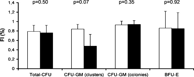
Purpose: We investigated the in vitro toxicity of bendamustine and fludarabine to hematopoietic progenitors and stem cells from healthy donors.
Methods: Clonogenic agar colony assays, non-clonogenic long-term liquid cultures (LTC) and apoptosis assays were used to assess the cytotoxicity of both the agents.
Results: Total colony-forming units (CFU) were more sensitive to fludarabine than to bendamustine in agar colony assays (IC(50) 0.7 microM/L and 8.5 microM/L, respectively). Using the Bliss independence model and combining the two agents yielded additive inhibition of progenitors. Non-clonogenic assays, including LTC and an apoptosis assay detecting activated caspases showed that stem cells are characterized by low sensitivity to bendamustine. In contrast, fludarabine strongly inhibited the viability and growth of stem cells in LTC.
Conclusions: Our data show that bendamustine is characterized by lower in vitro toxicity to hematopoietic progenitors and stem cells than fludarabine and might thus be preferable in regimens prior to stem cells apheresis.
Figures


References
-
- Adkins JC, Peters DH, Markham A (1997) Fludarabine. An update of its pharmacology and use in the treatment of haematological malignancies. Drugs 53:1005–1037 - PubMed
-
- Balfour JA, Goa KL (2001) Bendamustine. Drugs 61:631–640 - PubMed
-
- Blagosklonny MV (2004) Prospective strategies to enforce selectively cell death in cancer cells. Oncogene 23:2967–2975 - PubMed
-
- Bornhauser M, Storer B, Slattery JT, Appelbaum FR, Deeg HJ, Hansen J, Martin PJ, McDonald GB, Nichols WG, Radich J, Woolfrey A, Jenke A, Schleyer E, Thiede C, Ehninger G, Anasetti C (2003) Conditioning with fludarabine and targeted busulfan for transplantation of allogeneic hematopoietic stem cells. Blood 102:820–826 - PubMed
-
- Bröker LE, Huisman C, Ferreira CG, Rodriguez JA, Kruyt FA, Giaccone G (2002) Late activation of apoptotic pathways plays a negligible role in mediating the cytotoxic effects of discodermolide and epothilone B in non-small cell lung cancer cells. Cancer Res 62:4081–4088 - PubMed
Publication types
MeSH terms
Substances
LinkOut - more resources
Full Text Sources
Medical
