Review
. 2009 Jan;16(1):43-57.
doi: 10.1080/10739680802273892.

Neutrophil recruitment under shear flow: it's all about endothelial cell rings and gaps

Affiliations
Review

Neutrophil recruitment under shear flow: it's all about endothelial cell rings and gaps

Pilar Alcaide et al. Microcirculation. 2009 Jan.

Erratum in

  • Microcirculation. 2009 Nov;16(8):782

Abstract

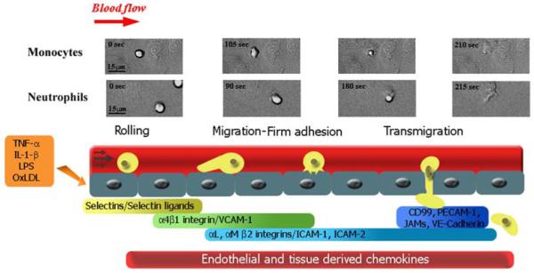
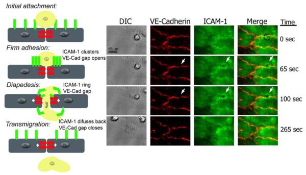
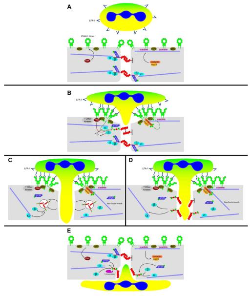
Leukocyte recruitment to tissues and organs is an essential component of host defense. The molecular mechanisms controlling this process are complex and remain under active investigation. The combination of biochemical techniques and live cell imaging using in vivo and in vitro flow-model approaches have shed light on several aspects of neutrophil transmigration through the vascular endothelial lining of blood vessels. Here, we focus on the role of adhesion molecule signaling in endothelial cells and their downstream targets during the process of transendothelial migration at cell-cell borders (paracellular transmigration). An emerging model involves the leukocyte beta2 integrin engagement of endothelial cell ICAM-1, which triggers integrin-ICAM-1 clustering (rings) and stabilizes leukocyte adhesion at cell-cell junctions. This step recruits nonreceptor tyrosine kinases that phosphorylate key tyrosine residues in the cytoplasmic tail of VE-cadherin, which destabilizes its linkage to catenins and the actin cytoskeleton, triggering the transient opening of VE-cadherin homodimers to form a gap in the cell junction, through which the neutrophil transmigrates. Interestingly, the signaling events that lead to neutrophil transmigration occur independently of shear flow in vitro.


Figures

Fig legend 1
Fig legend 1. Sequence of steps during leukocyte recruitment culminating in transendothelial migration
Circulating monocytes and neutrophils in the bloodstream initially attach to activated endothelium by selectin-mediated and α4β1 integrin-mediated interactions involved in the initial rolling step. The leukocytes locomote on the endothelial cell vessel wall by establishing Mac-1 integrin-mediated interactions with ICAM-1 on the endothelial cells until they firmly arrest near cell-cell junctions. This step is followed by stronger interactions mediated by several endothelial cell molecules besides ICAM-1, in which the leukocyte squeezes through the endothelial cell junction. Frames corresponding to live cell imaging digital microscopy show monocytes and neutrophils interacting with 4-hr TNF-α-activated HUVEC monolayers under flow conditions in vitro.
Figure legend 2
Figure legend 2. Live imaging of endothelial cell VE-Cadherin gaps and ICAM-1 rings dynamics during transmigration
HUVECs were activated with TNF-α for 4 h and stained with nonblocking mAbs to VE-Cadherin and ICAM-1 directly conjugated in red (Alexa 568) and green (Alexa 488), respectively. Freshly isolated neutrophils were drawn through a parallel plate flow chamber apparatus at 1 dyne/cm2 of shear stress. Three-channel, live-cell digital microscopy of neutrophils and endothelium during the process of transmigration was carried out as described previously (69). At time= 0, the neutrophil approaches the brightly stained cell junction during the initial attachment to the 4-hr TNF-α activated HUVECs. The dynamic behavior of VE-Cadherin and ICAM-1 has not started. At time=65 sec, the neutrophil firmly adheres to the endothelial cells at the junctions, and as VE-Cadherin starts forming a de novo gap, ICAM-1 clusters at the cell-cell junction where the neutrophil is firmly adhered. At t= 100 sec, the neutrophil is undergoing diapedesis, and part of it is still on top of the monolayer, while part of it is underneath. At this point the VE-Cadherin gap reaches its maximum aperture, and ICAM-1 forms a ring-like structure surrounding the transmigrating neutrophil. Finally, at t= 265 sec, the neutrophil has completely transmigrated, and the endothelial cell junction returns to normal: the VE-Cadherin gap is closed, and ICAM-1 is again uniformly distributed on the apical surface of the endothelial cell.
Figure 3
Figure 3. Outside-in signaling resulting in leukocyte transmigration at the junctions through activated endothelium: Interplay of endothelial cell molecules that participate actively in the dynamic reorganization of the adherens junctions
A: Pretransmigratory state. Endothelial cells express ICAM-1 in the closed “O-form” dimer, homogeneously distributed in the cytoplasmic membrane (with slight but detectable enrichment at cell-cell junctions). In the cytoplasm, ICAM-1 is linked to actin filaments via α-actinin. VE-cadherin is shown as transdimers, forming the adherens junction. VE-cadherin is linked to the actin cytoskeleton by α-, β-, and γ-catenin (plakoglobin). For clarity, γ-catenin is not included in the figure, but it is anticipated to behave in a similar manner, although this has not been investigated in great detail (4,5). p120-catenin binds to the juxtamembrane region in the VE-cadherin tail and does not function in linkage to the cytoskeleton; rather it functions in VE-cadherin retention on the surface(93). Protein tyrosine kinases (src, pyk2) and serine kinases (PAK) participate in the induction of cytoskeletal remodeling after ICAM-1 engagement (3,98). Early events in leukocyte transmigration will include ICAM-1 clustering and consequent recruitment of cortactin-Arp2/3 and protein kinases to the clustered ICAM-1 tails. Others have shown that Rho G and its GEF, SEGF, also bind to the ICAM-1 cytoplasmic tail at this step in transmigration (82). A leukocyte is depicted with LFA-1 in the inactive state. B: Initial stage of leukocyte transmigration. Engagement of ICAM-1 by leukocyte LFA-1 (in the active conformation) causes ICAM-1 clustering, and we suggest that a conformational switch occurs from the O-form to the W-form based on recent evidence from Yang and colleagues (99). Within the endothelial cell cytoplasm, protein kinases recruited to clustered ICAM-1 tails become phosphorylated and catalyze the phosphorylation of cortactin, VE-cadherin, and p120. Green arrows depict tyrosine phosphorylation, while PAK-catalyzed phosphorylation of VE-cadherin ser665 (80) is represented by a red arrow. The localization of PAK is not known, but the presence of a proline-rich SH3 binding region in PAK (11) suggests that it associates with Src kinase. As a result of VE-cadherin phosphorylation, the catenin complex bound to VE-cadherin begins to disassemble, uncoupling VE-cadherin from the actin cytoskeleton. The adherens junction itself begins to dissociate as well. Cortactin, once phosphorylated, can serve as a scaffold for new actin branches (depicted in panels C and D below). C and D: Intermediate stage of leukocyte transmigration. Panel C illustrates the possibility that phosphorylated VE-cadherin is internalized within clathrin-coated vesicles (28,42,93,94) during leukocyte diapedesis, while panel D depicts VE-cadherin as remaining junctional but no longer participating in transdimerization with neighboring cells. The speed with which leukocyte diapedesis takes place suggests that the latter model is more likely than the former. This is pure speculation at this point. E: Leukocyte transmigration nearly complete. The endothelial cell monolayer, having accommodated leukocyte transmigration, must reseal junctional gaps. This process is likely to involve protein tyrosine and serine phosphatases (whose identities have yet to be determined), which restore proteins to their resting states. VE-cadherin reassociates with the actin cytoskeleton, while actin branches formed by cortactin-Arp2/3 near the apical surface of the endothelial cell have dissolved.

References

    1. Adamson P, Etienne S, Couraud PO, Calder V, Greenwood J. Lymphocyte migration through brain endothelial cell monolayers involves signaling through endothelial ICAM-1 via a rho-dependent pathway. J Immunol. 1999;162:2964–2973. - PubMed
    1. Albelda SM, Muller WA, Buck CA, Newman PJ. Molecular and cellular properties of PECAM-1 (endoCAM/CD31): a novel vascular cell-cell adhesion molecule. J Cell Biol. 1991;114:1059–1068. - PMC - PubMed
    1. Allingham MJ, van Buul JD, Burridge K. ICAM-1-Mediated, Src- and Pyk2-Dependent Vascular Endothelial Cadherin Tyrosine Phosphorylation Is Required for Leukocyte Transendothelial Migration. J Immunol. 2007;179:4053–4064. - PubMed
    1. Allport JR, Ding H, Collins T, Gerritsen ME, Luscinskas FW. Endothelial-dependent mechanisms regulate leukocyte transmigration: A process involving the proteasome and disruption of the vascular endothelial-cadherin complex at endothelial cell-to-cell junctions. J Exp Med. 1997;186:517–527. - PMC - PubMed
    1. Allport JR, Muller WA, Luscinskas FW. Monocytes induce reversible focal changes in vascular endothelial cadherin complex during transendothelial migration under flow. J Cell Biol. 2000;148:203–216. - PMC - PubMed

Publication types