

Optical coherence tomography of clear corneal incisions for cataract surgery
- PMID: 18721720
- PMCID: PMC2556292
- DOI: 10.1016/j.jcrs.2008.05.026
Optical coherence tomography of clear corneal incisions for cataract surgery
Abstract
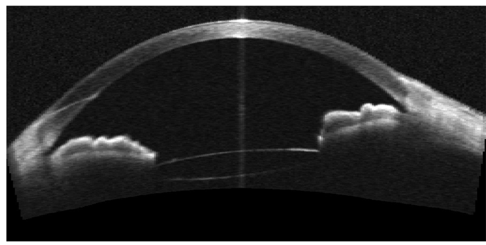
Purpose: To study the architecture of clear corneal incisions for phacoemulsification cataract surgery using optical coherence tomography (OCT).
Setting: Doheny Eye Institute and Department of Ophthalmology, Keck School of Medicine of the University of Southern California, Los Angeles, California, USA.
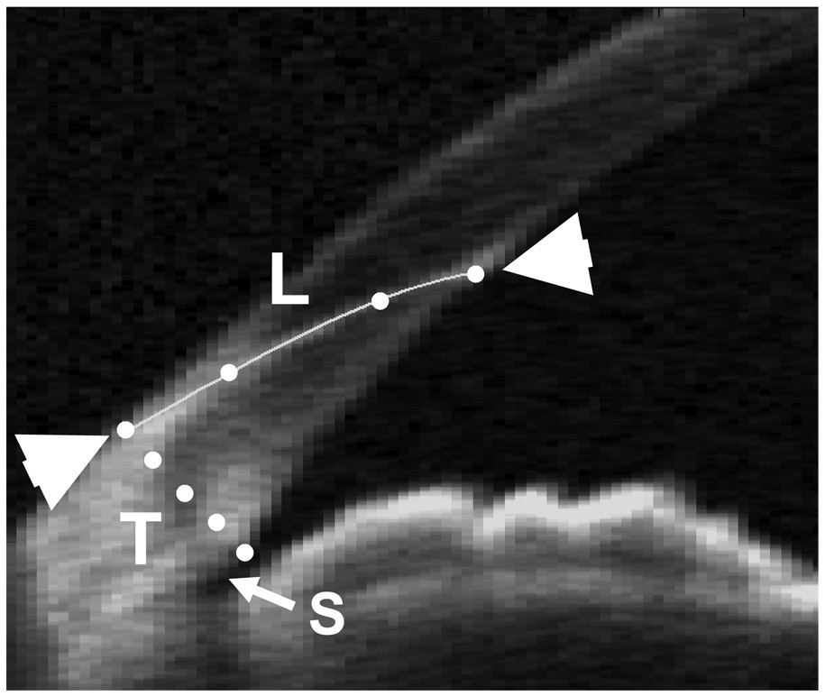
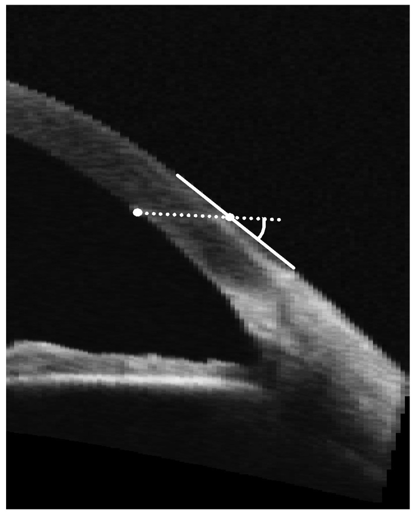
Methods: This prospective study comprised 20 eyes of 20 patients 1 month after cataract surgery performed by 1 of 2 experienced surgeons. Temporal clear corneal single-plane incisions were made with 3.0 mm metal keratomes. Each eye was scanned before and 1 month after surgery with a prototype high-speed anterior segment OCT system (Carl Zeiss Meditec, Inc.). The OCT scans were repeated 3 times during the same visit. The length of the corneal incision, thickness of the cornea, and position of the incision (distance from external wound edge to scleral spur) were measured using a computer caliper. The angle of the incision relative to the corneal surface was then calculated.
Results: The mean corneal incision length was 1.81 mm +/- 0.27 (SD), the mean corneal thickness at the incision was 747 +/- 67 microm, and the mean distance between the incision and the scleral spur was 1.46 +/- 0.24 mm. The mean angle of the incision was 26.8 +/- 5.5 degrees. The measurements were repeatable to within +/-0.072 mm (pooled standard deviation) for the incision length, +/-11 microm for the corneal thickness, and +/-0.042 mm for the position of the incision.
Conclusions: Optical coherence tomography allowed excellent evaluation of corneal incisions in cataract surgery postoperatively. Measurements of wound dimensions using OCT were highly repeatable.
Figures
References
-
- Nagaki Y, Hayasaka S, Kadoi C, et al. Bacterial endophthalmitis after small-incision cataract surgery. J Cataract Refract Surg. 2003;29:20–26. - PubMed
-
- Cooper BA, Holekamp NM, Bohigian G, Thompson PA. Case-control Study of Endophthalmitis After Cataract Surgery Comparing Scleral Tunnel and Clear Corneal Wounds. Am J Ophthalmol. 2003;136:300–305. - PubMed
-
- Taban M, Behrens A, Newcomb RL, et al. Acute Endophthalmitis Following Cataract Surgery. Arch Ophthalmol. 2005;123:613–620. - PubMed
-
- Mackool RJ, Russell RS. Strength of clear corneal incisions in cadaver eyes. J Cataract Refract Surg. 1996;22:721–725. - PubMed
-
- Taban M, Rao B, Reznik J, et al. Dynamic Morphology of Sutureless Cataract Wounds - Effect of Incision Angle and Location. Surv Ophthalmol. 2004;49 Suppl 2:S62–S72. - PubMed
