

Allergy and eosinophil-associated gastrointestinal disorders (EGID)
- PMID: 18721876
- PMCID: PMC2615814
- DOI: 10.1016/j.coi.2008.07.010
Allergy and eosinophil-associated gastrointestinal disorders (EGID)
Abstract
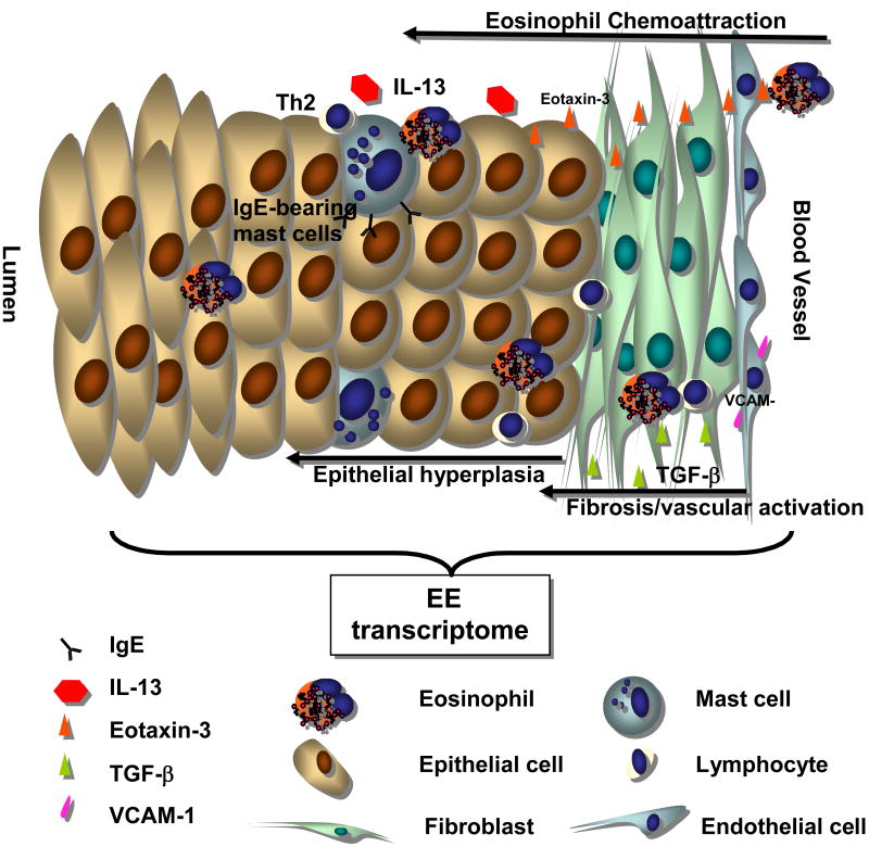
Eosinophil-associated gastrointestinal disorders (EGIDs) are characterized by an inappropriate accumulation of eosinophils within the gastrointestinal tract. The underlying etiology and pathophysiology that lead to the development of EGID are far from elucidated. However, there is growing evidence to support the role of aeroallergens and food allergens in the pathogenesis of these disorders. Recent advances have highlighted the role of Th2-driven cytokines in the development of EGID, and clinical studies have verified that children and adults with EGID often have positive skin testing to food allergens. The most common form of EGID, eosinophilic esophagitis (EE), has garnered intense investigation following an increased recognition over the past decade. Recently, there have been several important studies providing insight into both the cellular mechanisms governing EE and clinical therapies directed toward the treatment of EE. In the article herein, we will review the most recent scientific advances influencing our understanding of EGID with special emphasis on the role of allergens in the pathogenesis of EGID.
Figures
References
-
- DeBrosse CW, Case JW, Putnam PE, Collins MH, Rothenberg ME. Quantity and distribution of eosinophils in the gastrointestinal tract of children. Pediatr Dev Pathol. 2006;9:210–8. - PubMed
-
- Rothenberg ME, Mishra A, Brandt EB, Hogan SP. Gastrointestinal Eosinophils. Immunol Rev. 2001;179:139–55. - PubMed
-
- Noel RJ, Putnam PE, Rothenberg ME. Eosinophilic Esophagitis. N Engl J Med. 2004;351:1422–30. - PubMed
-
- Straumann A, Simon HU. Eosinophilic Esophagitis-escalating epidemiology? J Allergy Clin Immunol. 2005;115:418–9. - PubMed
-
- Putnam PE. Eosinophilic Esophagitis in Children: Clinical Manifestations. Gastrointest Endoscopy Clin N Am. 2008;18:11–23. - PubMed
Publication types
MeSH terms
Substances
Grants and funding
LinkOut - more resources
Full Text Sources
Other Literature Sources
Medical
Miscellaneous
