Achalasia: a new clinically relevant classification by high-resolution manometry
- PMID: 18722376
- PMCID: PMC2894987
- DOI: 10.1053/j.gastro.2008.07.022
Achalasia: a new clinically relevant classification by high-resolution manometry
Abstract
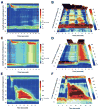
Background & aims: Although the diagnosis of achalasia hinges on demonstrating impaired esophagogastric junction (EGJ) relaxation and aperistalsis, 3 distinct patterns of aperistalsis are discernable with high-resolution manometry (HRM). This study aimed to compare the clinical characteristics and treatment response of these 3 subtypes.
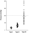
Methods: One thousand clinical HRM studies were reviewed, and 213 patients with impaired EGJ relaxation were identified. These were categorized into 4 groups: achalasia with minimal esophageal pressurization (type I, classic), achalasia with esophageal compression (type II), achalasia with spasm (type III), and functional obstruction with some preserved peristalsis. Clinical and manometric variables including treatment response were compared among the 3 achalasia subtypes. Logistic regression analysis was performed using treatment success as the dichotomous dependent variable controlling for independent manometric and clinical variables.
Results: Ninety-nine patients were newly diagnosed with achalasia (21 type I, 49 type II, 29 type III), and 83 of these had sufficient follow-up to analyze treatment response. Type II patients were significantly more likely to respond to any therapy (BoTox [71%], pneumatic dilation [91%], or Heller myotomy [100%]) than type I (56% overall) or type III (29% overall) patients. Logistic regression analysis found type II to be a predictor of positive treatment response, whereas type III and pretreatment esophageal dilatation were predictive of negative treatment response.
Conclusions: Achalasia can be categorized into 3 subtypes that are distinct in terms of their responsiveness to medical or surgical therapies. Utilizing these subclassifications would likely strengthen future prospective studies of treatment efficacy in achalasia.
Conflict of interest statement
Conflict of interests: John Pandolfino (consultant: Astra Zeneca, Santarus, Medtronic, Crospon, TAP; speaker: Astra Zeneca, Santarus, Medtronic); Monika Kwiatek (none), Thomas Nealis (none), William Bulsiewicz (none), Jennifer Post (none), and Peter J. Kahrilas (none). Other conflicts of interest were not disclosed because this was a retrospective analysis of medical records and manometry studies.
Figures


Comment in
-
The clinical usefulness of high-resolution manometry for the management of achalasia.Curr Gastroenterol Rep. 2009 Jun;11(3):170-2. doi: 10.1007/s11894-009-0027-1. Curr Gastroenterol Rep. 2009. PMID: 19463215 No abstract available.
-
[High-resolution manometry for classification of achalasia].Z Gastroenterol. 2010 Jun;48(6):709-10. doi: 10.1055/s-0029-1245290. Epub 2010 Jun 1. Z Gastroenterol. 2010. PMID: 20517810 German. No abstract available.
References
-
- Pandolfino JE, Kahrilas PJ. AGA technical review on the clinical use of esophageal manometry. Gastroenterology. 2005;128:209–224. - PubMed
-
- Goldenberg SP, Burrell M, Fette GG, et al. Classic and vigorous achalasia: a comparison of manometric, radiographic, and clinical findings. Gastroenterology. 1991;101:743–748. - PubMed
-
- Todorczuk JR, Aliperti G, Staiano A, et al. Reevaluation of manometric criteria for vigorous achalasia. Is this a distinct clinical disorder? Dig Dis Sci. 1991;36:274–278. - PubMed
-
- Hirano I, Tatum RP, Shi G, et al. Manometric heterogeneity in patients with idiopathic achalasia. Gastroenterology. 2001;120:789–798. - PubMed
Publication types
MeSH terms
Grants and funding
LinkOut - more resources
Full Text Sources
Medical

