

Metabolic control of T cell activation and death in SLE
- PMID: 18722557
- PMCID: PMC2680195
- DOI: 10.1016/j.autrev.2008.07.041
Metabolic control of T cell activation and death in SLE
Abstract
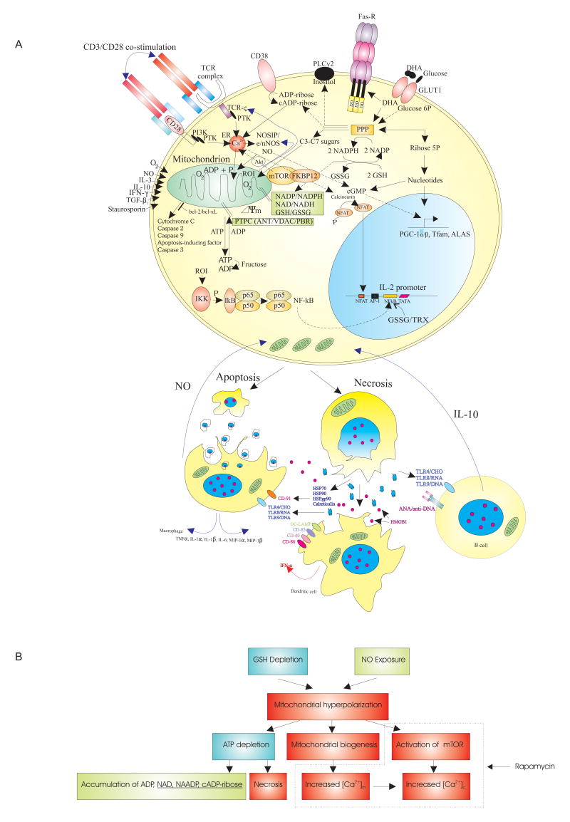
Systemic lupus erythematosus (SLE) is characterized by abnormal T cell activation and death, processes which are crucially dependent on the controlled production of reactive oxygen intermediates (ROI) and of ATP in mitochondria. The mitochondrial transmembrane potential (Deltapsi(m)) has conclusively emerged as a critical checkpoint of ATP synthesis and cell death. Lupus T cells exhibit persistent elevation of Deltapsi(m) or mitochondrial hyperpolarization (MHP) as well as depletion of ATP and glutathione which decrease activation-induced apoptosis and instead predispose T cells for necrosis, thus stimulating inflammation in SLE. NO-induced mitochondrial biogenesis in normal T cells accelerates the rapid phase and reduces the plateau of Ca(2+) influx upon CD3/CD28 co-stimulation, thus mimicking the Ca(2+) signaling profile of lupus T cells. Treatment of SLE patients with rapamycin improves disease activity, normalizes CD3/CD28-induced Ca(2+) fluxing but fails to affect MHP, suggesting that altered Ca(2+) fluxing is downstream or independent of mitochondrial dysfunction. Understanding the molecular basis and consequences of MHP is essential for controlling T cell activation and death signaling in SLE.
Figures
Similar articles
-
Nitric oxide-dependent mitochondrial biogenesis generates Ca2+ signaling profile of lupus T cells.J Immunol. 2004 Sep 15;173(6):3676-83. doi: 10.4049/jimmunol.173.6.3676. J Immunol. 2004. PMID: 15356113 Free PMC article.
-
Mitochondrial hyperpolarization and ATP depletion in patients with systemic lupus erythematosus.Arthritis Rheum. 2002 Jan;46(1):175-90. doi: 10.1002/1529-0131(200201)46:1<175::AID-ART10015>3.0.CO;2-H. Arthritis Rheum. 2002. PMID: 11817589 Free PMC article.
-
T cell activation-induced mitochondrial hyperpolarization is mediated by Ca2+- and redox-dependent production of nitric oxide.J Immunol. 2003 Nov 15;171(10):5188-97. doi: 10.4049/jimmunol.171.10.5188. J Immunol. 2003. PMID: 14607919 Free PMC article.
-
Mitochondrial dysfunction in T cells of patients with systemic lupus erythematosus.Int Rev Immunol. 2004 May-Aug;23(3-4):293-313. doi: 10.1080/08830180490452576. Int Rev Immunol. 2004. PMID: 15204090 Review.
-
The role of nitric oxide in abnormal T cell signal transduction in systemic lupus erythematosus.Clin Immunol. 2006 Feb-Mar;118(2-3):145-51. doi: 10.1016/j.clim.2005.10.016. Epub 2006 Jan 10. Clin Immunol. 2006. PMID: 16406340 Free PMC article. Review.
Cited by
-
Metabolic disturbances associated with systemic lupus erythematosus.PLoS One. 2012;7(6):e37210. doi: 10.1371/journal.pone.0037210. Epub 2012 Jun 19. PLoS One. 2012. PMID: 22723834 Free PMC article.
-
Increased mitochondrial electron transport chain activity at complex I is regulated by N-acetylcysteine in lymphocytes of patients with systemic lupus erythematosus.Antioxid Redox Signal. 2014 Jul 1;21(1):56-65. doi: 10.1089/ars.2013.5702. Epub 2014 Apr 23. Antioxid Redox Signal. 2014. PMID: 24673154 Free PMC article.
-
A cellular overview of immunometabolism in systemic lupus erythematosus.Oxf Open Immunol. 2023 May 11;4(1):iqad005. doi: 10.1093/oxfimm/iqad005. eCollection 2023. Oxf Open Immunol. 2023. PMID: 37554724 Free PMC article. Review.
-
Fus1/Tusc2 is a novel regulator of mitochondrial calcium handling, Ca2+-coupled mitochondrial processes, and Ca2+-dependent NFAT and NF-κB pathways in CD4+ T cells.Antioxid Redox Signal. 2014 Apr 1;20(10):1533-47. doi: 10.1089/ars.2013.5437. Epub 2014 Feb 4. Antioxid Redox Signal. 2014. PMID: 24328503 Free PMC article.
-
The Impact of Protein Acetylation/Deacetylation on Systemic Lupus Erythematosus.Int J Mol Sci. 2018 Dec 12;19(12):4007. doi: 10.3390/ijms19124007. Int J Mol Sci. 2018. PMID: 30545086 Free PMC article. Review.
References
-
- Kyttaris VC, Juang YT, Tsokos GC. Immune cells and cytokines in systemic lupus erythematosus: an update. Curr Opin Rheumatol. 2005;17:518–522. [see comment]. [Review] [29 refs] - PubMed
Publication types
MeSH terms
Substances
Grants and funding
LinkOut - more resources
Full Text Sources
Other Literature Sources
Medical
Miscellaneous
