Review
. 2009 Jan;1789(1):58-68.
doi: 10.1016/j.bbagrm.2008.07.009. Epub 2008 Aug 3.

Histone modifying enzymes: structures, mechanisms, and specificities

Affiliations
Review

Histone modifying enzymes: structures, mechanisms, and specificities

Ronen Marmorstein et al. Biochim Biophys Acta. 2009 Jan.

Abstract

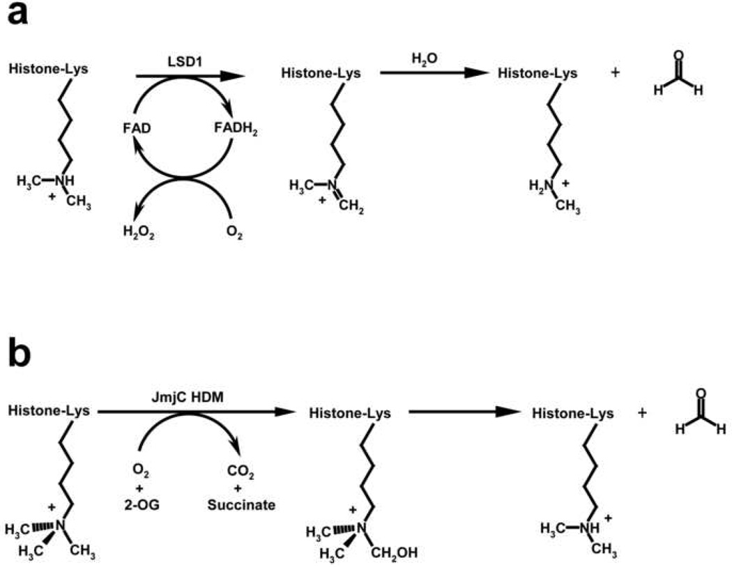
Histone modifying enzymes catalyze the addition or removal of an array of covalent modifications in histone and non-histone proteins. Within the context of chromatin, these modifications regulate gene expression as well as other genomic functions and have been implicated in establishing and maintaining a heritable epigenetic code that contributes to defining cell identity and fate. Biochemical and structural characterization of histone modifying enzymes has yielded important insights into their respective catalytic mechanisms, substrate specificities, and regulation. In this review, we summarize recent advances in understanding these enzymes, highlighting studies of the histone acetyltransferases (HATs) p300 (also now known as KAT3B) and Rtt109 (KAT11) and the histone lysine demethylases (HDMs) LSD1 (KDM1) and JMJD2A (KDM4A), present overriding themes that derive from these studies, and pose remaining questions concerning their regulatory roles in mediating DNA transactions.


Figures

Figure 1
Figure 1. Overall structure of nuclear HATs
(a) Tetrahymena Gcn5 (1PUA.pdb) is shown in complex with CoA (stick representation in CPK coloring) and histone H3 (backbone representation in green coloring) illustrating a representative Gcn5/PCAF HAT. The protein is shown in cartoon representation with the structurally conserved HAT core region highlighted in blue and the more structurally variable HAT regions shown in cyan. (b) Yeast Esa1 in complex with CoA (1FY7.pdb) illustrates a representative MYST HAT. The coloring is as described in part (a). (c) Human p300 in complex with the Lys-CoA bisubstrate inhibitor (3BIY.pdb) shows a representative p300/CBP HAT. The coloring is as described in part (a) and the substrate binding loop is highlighted in cartoon representation in red. (d) Yeast Rtt109 in complex with Ac-CoA (3D35.pdb). The coloring is as described in part (c).
Figure 2
Figure 2. p300/CBP substrate binding site and enzymatic mechanism
(a) The histone/protein binding site groove is highlighted. An electrostatic surface of p300 is shown with positive, negative, and neutral charged surfaces shown in blue, red, and white, respectively. The two pronounced electronegative p300 pockets that are proposed to participate in protein substrate binding are highlighted with black dotted circles. Lys-CoA is shown in yellow stick representation. (b) The active site highlighting residues (CPK coloring with carbon atoms in green) that are in position to play catalytic roles. (c) The Theorell-Chance (hit and run) catalytic mechanism is shown. (d) A model for regulation of p300/CBP by autoacetylation is shown where it is proposed that the lysine-rich basic activation loop blocks the histone/protein binding site in the hypoacetylated form and is released from this site upon autoacetylation.
Figure 3
Figure 3. Structural details of the Rtt109/Ac-CoA complex
(a) The active site highlighting residues that are mutationally sensitive for HAT activity. Ac-CoA is shown in CPK coloring and stick representation. (b) Autoacetylated Lys290 is shown hydrogen bonded to Asp288 and buried within a hydrophobic core of phenylalanine residues.
Figure 4
Figure 4. Chemical mechanisms of lysine demethylation
(a) FAD-dependent demethylation of mono- and dimethyllysines by LSD1. The oxidation of the methyl ε-ammonium group forms an imine intermediate that subsequently hydrolyzes to yield the demethylated lysine and a molecule of formaldehyde. (b) Fe(II)-dependent demethylation of mono-, di-, and trimethyllysines by JmjC HDMs. Using the co-substrates O2 and 2-OG, JmjC enzymes catalyze the hydroxylation of a lysine methyl group via a radical-based mechanism, forming the products succinate and carbon dioxide. The hydroxyl-methyl ε-ammonium hemiaminal intermediate subsequently decomposes to yield the demethylated lysine and formaldehyde.
Figure 5
Figure 5. Structural basis for the substrate specificity of LSD1
(a) Crystal structure of a human LSD1/Co-REST complex covalently bound to a histone H3 peptide suicide inhibitor (2UXN.pdb). The secondary structure of the SWIRM (blue), AOD (gray), and Tower domains of LSD1 and the linker (magenta) and SANT2 (red) domains of Co-REST are illustrated in cartoon representation. The covalently modified FAD cofactor and H3K4 inhibitor are shown in yellow stick representation. (b) Structure of LSD1’s active site bound to the H3 peptide methylpropargyl-K4 (K4mp) inhibitor that forms a covalent adduct with FAD (CPK coloring with yellow carbon atoms). Residues in LSD1 that interact with the H3 peptide are shown and labeled in black. Hydrogen bonds are delineated as orange dashed lines. (c) Structure of LSD1/Co-REST complex bound to an H3K4M peptide inhibitor (cyan carbons) (2V1D.pdb) illustrated as in (b). The C-terminal residues K14, A15, and P16 in the H3 peptide are omitted for clarity. (d) Superimposition of the H3 peptide inhibitors in (b) and (c) illustrating their distinct binding conformations within the substrate binding cleft of LSD1.
Figure 6
Figure 6. Dual specificity of JMJD2A for H3K9me3 and H3K36me3
(a) Crystal structure of the catalytic domain of human JMJD2A bound to an H3K9me3 peptide (2OQ6.pdb). The JmjN domain (blue), mixed region (magenta), JmjC domain (gray), and C-terminal region (green) that comprise the catalytic domain are shown in cartoon representation. The H3K9me3 peptide (CPK coloring with cyan carbon atoms) and NOG (yellow carbons) are rendered in stick representation. The His188–Glu190–His276 triad that coordinates the active site Ni(II) (teal) and the Zn atom (dark gray) that forms the Zn-finger motif between the JmjC domain and C-terminal region are also illustrated. (b) Substrate binding cleft of JMJD2A bound to the H3K9me3 peptide, NOG, and Ni(II). Residues in JMJD2A that interact with the peptide are colored according to their domain association (a) and labeled in black. Hydrogen bonds are shown as in Figure 4b. (c) Structure of JMJD2A bound to an H3K36me3 peptide (orange carbons) (2OS2.pdb) illustrated as in (b). Residues H39 and R40 in the H3 peptide are modeled as alanines owing to the ambiguous electron density observed for their side chains. (d) Superimposition of the H3K9me3 and H3K36me3 peptides bound to JMJD2A, illuminating the similarities and differences in the binding conformations of the two substrates.
Figure 7
Figure 7. Methylation state specificity of JMJD2A
(a) Structure of the active center of JMJD2A bound to K9me3 (CPK coloring with cyan carbon atoms), Ni(II) (teal), and NOG (yellow carbons) (2Q8C.pdb). Key residues that engage in CH⋯O hydrogen bonds with the methyllysine substrates are delineated. The dashed red line denotes the reaction coordinate between the ζ-methyl group positioned for hydroxylation and the water molecule bound in the presumptive O2 coordination site. (b) The active site of JMJD2A in complex with K9me2 (2OX0.pdb) illustrated as in (a). The alternative conformation of dimethyllysine side chain is shown with pink carbon atoms. (c) The active site of JMJD2A bound to K9me1 with its methyl group oriented distal from the metal center (2OT7.pdb), precluding hydroxylation and demethylation.

References

    1. Luger K, Hansen JC. Nucleosome and chromatin fiber dynamics. Curr Opin Struct Biol. 2005;15:188–196. - PubMed
    1. Kouzarides T. Chromatin modifications and their function. Cell. 2007;128:693–705. - PubMed
    1. Allfrey VG, Faulkner R, Mirsky AE. Acetylation and Methylation of Histones and Their Possible Role in the Regulation of Rna Synthesis. Proc Natl Acad Sci U S A. 1964;51:786–794. - PMC - PubMed
    1. Murray K. The Occurrence of Epsilon-N-Methyl Lysine in Histones. Biochemistry. 1964;3:10–15. - PubMed
    1. Kleinsmith LJ, Allfrey VG, Mirsky AE. Phosphoprotein metabolism in isolated lymphocyte nuclei. Proc Natl Acad Sci U S A. 1966;55:1182–1189. - PMC - PubMed

Publication types

MeSH terms