

Drusen ultrastructure imaging with spectral domain optical coherence tomography in age-related macular degeneration
- PMID: 18722666
- PMCID: PMC3510676
- DOI: 10.1016/j.ophtha.2008.04.041
Drusen ultrastructure imaging with spectral domain optical coherence tomography in age-related macular degeneration
Abstract
Purpose: To categorize drusen ultrastructure in age-related macular degeneration (AMD) using spectral domain optical coherence tomography (SDOCT) and correlate the tomographic and photographic drusen appearances.
Design: Prospective case series.
Participants: Thirty-one eyes of 31 patients with non-neovascular AMD.
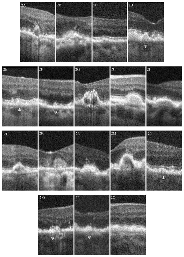
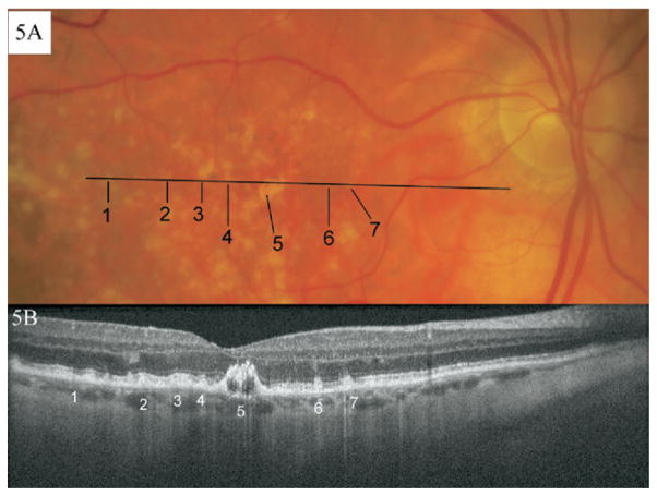
Methods: Subjects with drusen and a clinical diagnosis of AMD were enrolled in an SDOCT imaging study from August of 2005 to May of 2007. Foveal linear scans were acquired, and the image data were processed for analysis. Drusen were scored by 4 morphologic categories: shape, predominant internal reflectivity, homogeneity, and presence of overlying hyper-reflective foci. The prevalences of each morphologic pattern and combinations of morphologic patterns observed were calculated. The photographic appearance of each druse was compared with the tomographic classification. Interobserver and intraobserver agreement analysis was performed.
Main outcome measures: Prevalence of morphologic parameters using SDOCT.
Results: Twenty-one eyes of 21 patients had SDOCT B-scans of adequate quality for analysis. On the basis of the above morphologic categories, 17 different drusen patterns were found in 120 total drusen. The most common was convex, homogeneous, with medium internal reflectivity, and without overlying hyper-reflective foci, present in 17 of 21 eyes (81%). Of the 16 eyes (76%) with nonhomogeneous drusen, 5 had a distinct hyper-reflective core. Hyper-reflective foci overlying drusen were in 7 eyes (33%). Although half of the photographically soft-indistinct drusen were convex with medium internal reflectivity and homogeneous without overlying hyper-reflective foci, the other half had significant variability in their tomographic appearance. Both interobserver and intraobserver agreement in drusen grading were high. Readers agreed the most when grading drusen shape and reflectivity, whereas the least agreement was for drusen homogeneity.
Conclusions: Drusen ultrastructure can be imaged with SDOCT and characterized with a simple grading system. Photographic appearance may predict some but not all tomographic appearances. Trained observers have a high level of agreement with this grading system. These in vivo morphologic characteristics imaged with SDOCT may be distinct subclasses of drusen types, may relate closely to ultrastructural drusen elements identified in cadaveric eyes, and may be useful imaging biomarkers for disease severity or risk of progression. This will require validation from further studies.
Figures


References
-
- Anderson DH, Talaga KC, Rivest AJ, et al. Characterization of beta amyloid assemblies in drusen: the deposits associated with aging and age-related macular degeneration. Exp Eye Res. 2004;78:243–56. - PubMed
-
- Anderson DH, Mullins RF, Hageman GS, Johnson LV. A role for local inflammation in the formation of drusen in the aging eye. Am J Ophthalmol. 2002;134:411–31. - PubMed
-
- Hageman GS, Luthert PJ, Victor Chong NH, et al. An integrated hypothesis that considers drusen as biomarkers of immune-mediated processes at the RPE-Bruch’s membrane interface in aging and age-related macular degeneration. Prog Retin Eye Res. 2001;20:705–32. - PubMed
-
- Klein R, Davis MD, Magli YL, et al. The Wisconsin Age-Related Maculopathy Grading System. Ophthalmology. 1991;98:1128–34. - PubMed
-
- Age-Related Eye Disease Study Research Group. The Age-Related Eye Disease Study system for classifying age-related macular degeneration from stereoscopic color fundus photographs: the Age-Related Eye Disease Study report number 6. Am J Ophthalmol. 2001;132:668– 81. - PubMed
Publication types
MeSH terms
Grants and funding
LinkOut - more resources
Full Text Sources
Other Literature Sources
Medical
