. 2008 Nov 14;383(3):713-30.
doi: 10.1016/j.jmb.2008.08.017. Epub 2008 Aug 13.

Organization and evolution of the biological response to singlet oxygen stress

Affiliations

Organization and evolution of the biological response to singlet oxygen stress

Yann S Dufour et al. J Mol Biol. .

Abstract

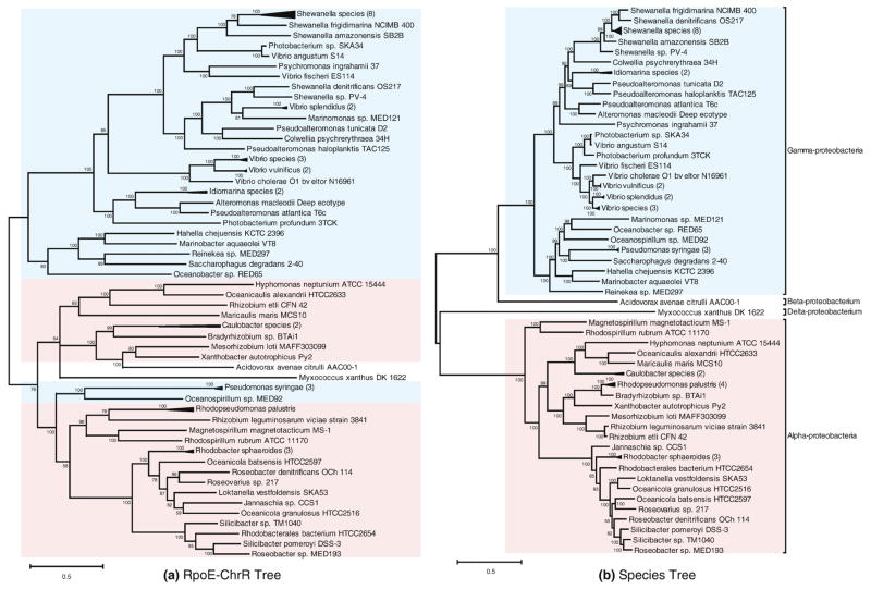
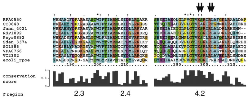
The appearance of atmospheric oxygen from photosynthetic activity led to the evolution of aerobic respiration and responses to the resulting reactive oxygen species. In Rhodobacter sphaeroides, a photosynthetic alpha-proteobacterium, a transcriptional response to the reactive oxygen species singlet oxygen ((1)O(2)) is controlled by the group IV sigma factor sigma(E) and the anti-sigma factor ChrR. In this study, we integrated various large datasets to identify genes within the (1)O(2) stress response that contain sigma(E)-dependent promoters both within R. sphaeroides and across the bacterial phylogeny. Transcript pattern clustering and a sigma(E)-binding sequence model were used to predict candidate promoters that respond to (1)O(2) stress in R. sphaeroides. These candidate promoters were experimentally validated to nine R. sphaeroides sigma(E)-dependent promoters that control the transcription of 15 (1)O(2)-activated genes. Knowledge of the R. sphaeroides response to (1)O(2) and its regulator sigma(E)-ChrR was combined with large-scale phylogenetic and sequence analyses to predict the existence of a core set of approximately eight conserved sigma(E)-dependent genes in alpha-proteobacteria and gamma-proteobacteria. The bacteria predicted to contain this conserved response to (1)O(2) include photosynthetic species, as well as free-living and symbiotic/pathogenic nonphotosynthetic species. Our analysis also predicts that the response to (1)O(2) evolved within the time frame of the accumulation of atmospheric molecular oxygen on this planet.


Figures

Fig. 1
Fig. 1
Expression profiles of σE–ChrR regulon members. Coregulated genes identified from the hierarchical clustering of genes differentially expressed in response to increased σE activity (ΔchrR mutant) compared to wild-type cells. Colors represent the relative abundance of transcripts in different conditions. Red indicates high expression level and green indicates low expression level, compared to the mean expression level. Genes are identified by their locus ID number. Brackets denote known or predicted transcriptional units.
Fig. 2
Fig. 2
Sequence logos of the predicted σE promoter motif in R. sphaeroides (a) and other bacteria (b). (a) The −35 and −10 motif logos were derived by sequence alignment of six σE-dependent promoters from R. sphaeroides. The two conserved regions are separated by a spacer of 13–14 bp. The information content (Iseq) of each motif is indicated. (b) The indicated −35 and −10 motifs were found upstream of the rpoE gene in 57 of the 73 selected microbial genomes. In both panels, the logos were produced with WebLogo [http://weblogo.berkeley.edu/].
Fig. 3
Fig. 3
In vitro transcription of target promoters using reconstituted R. sphaeroidesE. (a) Transcripts derived from the known σE-dependent promoter RSP1092 (rpoE) and a promoter identified in this study (RSP1852) in the presence of σE and ChrR. (b) Transcripts derived from RSP6222 and RSP3336 as a function of increasing σE levels. The amount of σE used in (b) is indicated relative to the amount used in the reaction shown in (a).
Fig. 4
Fig. 4
Activity of σE-dependent promoters in R. sphaeroides. Promoter activity is reported as the level of β-galatosidase activity (Miller units) in wild-type (gray), ΔchrR (black), and ΔrpoEchrR (white) cells. All assays were performed in triplicate, with bars representing standard deviations.
Fig. 5
Fig. 5
Localization of σE-dependent promoters by ChIP-chip. Shown are three representative genomic regions enriched by the immunoprecipitation of DNA fragments using anti-σE antibodies (red) or anti-β′ antibodies to assess RNA polymerase occupancy (blue). The data plot the log2 of the ratio of the immunoprecipitated sample to the control sample as a function of probe location along the R. sphaeroides genome (coordinates are indicated in base pairs). Regions significantly enriched by anti-σE immunoprecipitation (p≤0.01) are indicated by red blocks. The locations of the annotated open reading frames are indicated by black blocks (read forward or reverse if above or below the baseline, respectively). The positions and orientations of the known or predicted σE promoter are indicated by a green arrow. The data were plotted using SignalMap v1.9 (NimbleGen Systems).
Fig. 6
Fig. 6
Phylogeny of the σE–ChrR pair. Phylogenetic tree constructed with the concatenated amino acid sequence of σE and ChrR (a) or RuvB, RpoD, and GyrB (b) using Bayesian inference. Proteobacteria classes are indicated by color (pale blue for γ-proteobacteria and salmon for α-proteobacteria). Numbers on branches indicate Bayesian posterior probabilities. Branches of the tree for closely related species were collapsed for clarity.
Fig. 7
Fig. 7
Alignments of the amino acid sequences of σE homologs. Alignment of the regions 2.3–2.4 and 4.2 of σE homologs from a set of representative species containing σE–ChrR proteins. The histogram below the alignment represents the relative conservation score for each position based on alignment of all 73 σE homologs considered in this study. Arrows indicate residues predicted to contact DNA in the α35 region of the promoter based on alignment with E. coli σE. Sequences are identified by their locus ID.
Fig. 8
Fig. 8
Potential σE target genes across selected bacteria. (a) Groups of orthologous genes (columns) that contain the putative σE binding motif in their promoter regions across the indicated α-proteobacteria. (b) Same as in (a), but for the indicated γ-proteobacteria. A gray box means that the organism does not possess a homolog for the corresponding group; black means that it possesses a homolog for the group but no conserved σE binding motif was found in its promoter region; yellow means that a σE binding motif was found in the promoter region for the homologous gene. Shades of yellow represent the degree to which the gene is connected to the σE binding motif (see the text). A description and an annotation of these orthologs are presented in Table 3 and Supplemental Table 2.

References

    1. Kotelnikova EA, Makeev VJ, Gelfand MS. Evolution of transcription factor DNA binding sites. Gene. 2005;347:255–263. - PubMed
    1. Rhodius VA, Suh WC, Nonaka G, West J, Gross CA. Conserved and variable functions of the sigmaE stress response in related genomes. PLoS Biol. 2006;4:e2. - PMC - PubMed
    1. Koonin EV. Orthologs, paralogs, and evolutionary genomics. Annu Rev Genet. 2005;39:309–338. - PubMed
    1. Madan Babu M, Teichmann SA, Aravind L. Evolutionary dynamics of prokaryotic transcriptional regulatory networks. J Mol Biol. 2006;358:614–633. - PubMed
    1. Mackenzie C, Eraso JM, Choudhary M, Roh JH, Zeng X, Bruscella P, et al. Postgenomic adventures with Rhodobacter sphaeroides. Annu Rev Microbiol. 2007;61:283–307. - PubMed

Publication types

MeSH terms