Review
. 2008 Oct 31;283(44):29639-43.
doi: 10.1074/jbc.R800016200. Epub 2008 Aug 22.

Structural classification of toxic amyloid oligomers

Affiliations
Review

Structural classification of toxic amyloid oligomers

Charles G Glabe. J Biol Chem. .

Abstract

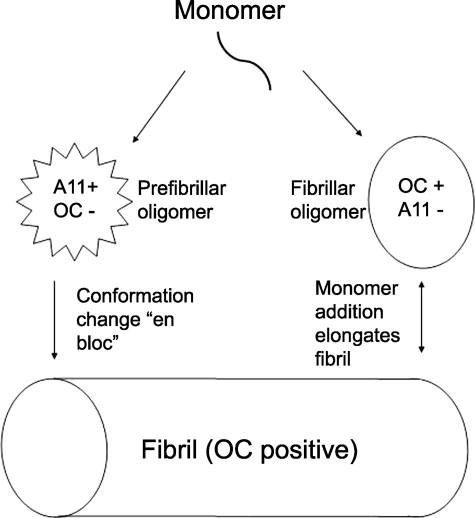
Amyloid oligomers are believed to play important causal roles in many types of amyloid-related degenerative diseases. Many different laboratories have reported amyloid oligomers that differ in size, morphology, toxicity, and method of preparation or purification, raising the question of the structural relationships among these oligomer preparations. The structural plasticity that has been reported to occur in amyloids formed from the same protein sequence indicates that it is quite possible that different oligomer preparations may represent distinct structural variants. In view of the difficulty in determining the precise structure of amyloids, conformation- and epitope-specific antibodies may provide a facile means of classifying amyloid oligomer structures. Conformation-dependent antibodies that recognize generic epitopes that are specifically associated with distinct aggregation states of many different amyloid-forming sequences indicate that there are at least two fundamentally distinct types of amyloid oligomers: fibrillar and prefibrillar oligomers. Classification of amyloid oligomers according to their underlying structures may be a more useful and rational approach than relying on differences in size and morphology.


Figures

FIGURE 1.
FIGURE 1.
Schematic representation of the distinct types of amyloid oligomers and fibrils. The aggregation pathway begins with a misfolded amyloidogenic monomer (top) and can diverge into two paths depending on which conformation it adopts. Monomers can aggregate to form prefibrillar oligomers that are A11-positive and OC-negative (left pathway). These prefibrillar oligomers may then align to form protofibrils (not shown) and undergo a concerted conformation change “en bloc” to form fibrils. They are termed prefibrillar oligomers because they are transient intermediates that ultimately become fibrils. In the other pathway, amyloidogenic monomers aggregate to form a fibrillar conformation or lattice that is OC-positive and A11-negative (right pathway). These fibrillar oligomers may represent fibril nuclei or seeds that are aggregates capable of elongating by recruiting additional monomers at their ends. Addition of monomers ultimately elongates the fibrils to a size that satisfies an arbitrary definition of insolubility and would be recognized as fibrillar under the electron or atomic force microscope, although no conformational difference is apparent by antibody reactivity. Fibrils may be distinct from fibrillar oligomers on the basis of their content of multiple protofilaments (not shown), but this does not imply that a fundamental conformation change in their integral peptide-building blocks is necessary for fibrillar oligomers to convert to fibrils. They may simply coalesce or grow by monomer addition to form fibrils.

References

    1. Kirschner, D. A., Abraham, C., and Selkoe, D. J. (1986) Proc. Natl. Acad. Sci. U. S. A. 83 503–507 - PMC - PubMed
    1. Wisniewski, H. M., and Terry, R. D. (1973) Prog. Neuropathol. 2 1–26
    1. Hardy, J., and Selkoe, D. J. (2002) Science 297 353–356 - PubMed
    1. Terry, R. D. (1996) J. Neuropathol. Exp. Neurol. 55 1023–1025 - PubMed
    1. Westerman, M. A., Cooper-Blacketer, D., Mariash, A., Kotilinek, L., Kawarabayashi, T., Younkin, L. H., Carlson, G. A., Younkin, S. G., and Ashe, K. H. (2002) J. Neurosci. 22 1858–1867 - PMC - PubMed

Publication types

LinkOut - more resources