. 2008 Oct 31;283(44):29929-37.
doi: 10.1074/jbc.M804065200. Epub 2008 Aug 22.

Hepatitis C viral NS3-4A protease activity is enhanced by the NS3 helicase

Affiliations

Hepatitis C viral NS3-4A protease activity is enhanced by the NS3 helicase

Rudolf K F Beran et al. J Biol Chem. .

Abstract

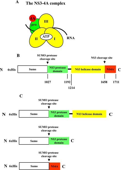
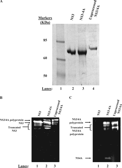
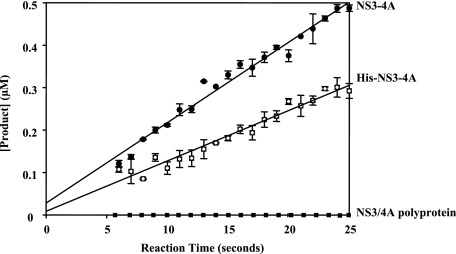
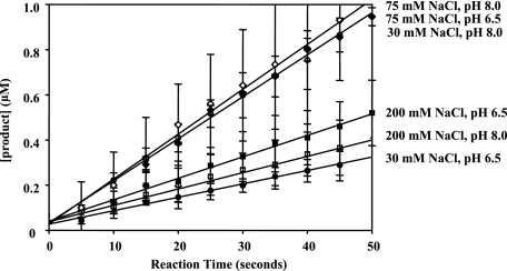
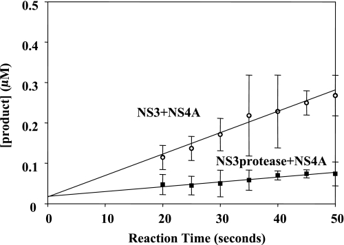
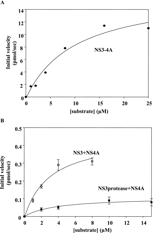
Non-structural protein 3 (NS3) is a multifunctional enzyme possessing serine protease, NTPase, and RNA unwinding activities that are required for hepatitis C viral (HCV) replication. HCV non-structural protein 4A (NS4A) binds to the N-terminal NS3 protease domain to stimulate NS3 serine protease activity. In addition, the NS3 protease domain enhances the RNA binding, ATPase, and RNA unwinding activities of the C-terminal NS3 helicase domain (NS3hel). To determine whether NS3hel enhances the NS3 serine protease activity, we purified truncated and full-length NS3-4A complexes and examined their serine protease activities under a variety of salt and pH conditions. Our results indicate that the helicase domain enhances serine protease activity, just as the protease domain enhances helicase activity. Thus, the two enzymatic domains of NS3-4A are highly interdependent. This is the first time that such a complete interdependence has been demonstrated for a multifunctional, single chain enzyme. NS3-4A domain interdependence has important implications for function during the viral lifecycle as well as for the design of inhibitor screens that target the NS3-4A protease.


Figures

FIGURE 1.
FIGURE 1.
Composition and purification of NS3-4A. The NS3-4A complex organization and construct design are illustrated schematically (A and B). In panel A, pro refers to the serine protease domain and the Roman numerals indicate the respective NS3 helicase subdomains. The regions where ATP, RNA, and the NS4A co-factor bind are indicated as well. The protein construct expressed in E. coli is depicted in panel B. The numbers below the map refer to the HCV polyprotein numbering of the amino acids of NS3-4A. In panel C, His-SUMO-NS3 and His-SUMO-NS4A fusion construct designs are presented. These proteins were purified separately and their His-SUMO tags were removed using the same methods as for NS3-4A. Subsequently, the native NS3 or native NS3 protease domain was reconstituted with native NS4A (see “Experimental Procedures”).
FIGURE 2.
FIGURE 2.
NS3-4A purifies as two separable proteins. A, purified proteins were subjected to denaturing electrophoresis on a 4–12% gradient gel. Purified NS3 (lane 2), NS3-4A (lane 3), and NS3/4A polyprotein (lane 4) were subjected to electrophoresis side by side for comparison of mobilities. The band shown in lane 3 represents the NS3 component of a native, fully cleaved NS3-4A preparation. The band shown in lane 4 represents an NS3/4A polyprotein preparation produced by mutating the Thr/Ser cleavage site between NS3 and NS4A to a non-cleavable sequence (AA). In panels B and C, anti-NS3 and anti-NS4A Western blot analysis confirm the identity of our purified proteins (see “Experimental Procedures”). Panel B depicts an anti-NS3 Western blot. In panel B, lane 1 contains purified NS3, lane 2 contains purified, full-length NS3-4A, and lane 3 contains purified NS3/4A polyprotein. Truncated forms of NS3 are visible below the full-length protein in each lane in the anti-NS3 blot. These truncated forms of NS3 are likely produced during bacterial expression as well as during the multiday purification performed in the absence of protease inhibitors. These truncated forms could represent either N- or C-terminal truncations of NS3 as the monoclonal anti-NS3 antibody binds to the central region of the helicase domain. Panel C depicts an anti-NS4A Western blot. In panel C, lane 1 contains purified NS3, lane 2 contains purified, full-length NS3-4A, and lane 3 contains purified NS3/4A polyprotein. Truncated forms of the NS3/4A polyprotein are visible below the full-length polyprotein in the anti-NS4A blot. These truncated forms of NS3/4A polyprotein likely represent N-terminal degraded NS3/4A as the monoclonal anti-NS4A antibody binds the final 11 C-terminal residues of NS4A.
FIGURE 3.
FIGURE 3.
Steady-state proteolysis of RET-S1 by NS3-4A and His-NS3-4A. The y intercept of the line in each case corresponds to the active fraction of protein (see “Experimental Procedures”). For NS3-4A (solid circles), the active fraction was 75 ± 14%. For His-NS3-4A (hatched squares), the active fraction was 25 ± 12%. NS3/4A polyprotein did not display any measurable serine protease activity (solid squares). The data shown are determined using proteins of the 1b genotype. Similar results were observed using NS3-4A(1a) and His-NS3-4A(1a). This data are the average of three experiments and the error values represent standard deviation.
FIGURE 4.
FIGURE 4.
Steady-state velocity curves for NS3-4A proteolysis of RET-S1 under a range of pH and salt conditions. The steady-state rates of proteolysis were 0.006 ± 0.001 μm product/s at pH 6.5, 30 mm NaCl (solid circles), 0.019 ± 0.001μm product/s at pH 8, 30 mm NaCl (pierced circles), 0.019 ± 0.001 μm product/s at pH 6.5, 75 mm NaCl (solid diamonds), 0.020 ± 0.001 μm product/s at pH 8, 75 mm NaCl (pierced diamonds), 0.010 ± 0.001 μm product/second at pH 6.5, 200 mm NaCl (solid squares), and 0.010 ± 0.001 μm product/s at pH 8, 200 mm NaCl (pierced squares). The active fraction in each case, as determined by intersection with the y intercept, was 68 ± 9% for pH 6.5, 30 mm NaCl, 84 ± 16% for pH 8.0, 30 mm NaCl, 84 ± 15% for pH 6.5, 75 mm NaCl, 70 ± 12% for pH 8.0, 75 mm NaCl, 95 ± 5% for pH 6.5, 200 mm NaCl, and 95 ± 5% for pH 8.0, 200 mm NaCl. The data shown were determined using NS3-4A of the 1b genotype and represent the steady-state data points fit to a line. Similar results were observed with NS3-4A (1a). This data are the average of three experiments and the error values represent standard deviation.
FIGURE 5.
FIGURE 5.
Steady-state proteolysis of RET-S1 by reconstituted NS3 + NS4A and NS3 protease domain + NS4A. The steady-state velocity for NS3 + NS4A is 0.005 ± 001 μm/s (pierced circles) and for NS3 protease ± NS4A is 0.001 ± 0.001 μm/s (solid squares). The y intercept of the fitted lines show NS3 + NS4A and NS3 protease domain + NS4A to have 75 ± 10% active fraction and 75 ± 12% active fraction, respectively. The data shown are the average of three experiments and the error values represent standard deviation.
FIGURE 6.
FIGURE 6.
Steady-state proteolysis rates of RET-S1 by NS3-4A, NS3 + NS4A, and NS3 protease domain + NS4A in the presence of varying substrate concentrations. The data were fit to the Michaelis-Menten equation to determine Km, Vmax, and kcat values. The data shown is the average of three experiments.

References

    1. Lam, A. M., and Frick, D. N. (2006) J. Virol. 80 404-411 - PMC - PubMed
    1. Lohmann, V., Korner, F., Koch, J., Herian, U., Theilmann, L., and Bartenschlager, R. (1999) Science 285 110-113 - PubMed
    1. de la Cruz, J., Kressler, D., and Linder, P. (1999) Trends Biochem. Sci. 24 192-198 - PubMed
    1. Failla, C., Tomei, L., and De Francesco, R. (1994) J. Virol. 68 3753-3760 - PMC - PubMed
    1. Taliani, M., Bianchi, E., Narjes, F., Fossatelli, M., Urbani, A., Steinkuhler, C., De Francesco, R., and Pessi, A. (1996) Anal. Biochem. 240 60-67 - PubMed

Publication types

MeSH terms