. 2008 Oct 24;283(43):29424-32.
doi: 10.1074/jbc.M802475200. Epub 2008 Aug 22.

Regulation of postsynaptic RapGAP SPAR by Polo-like kinase 2 and the SCFbeta-TRCP ubiquitin ligase in hippocampal neurons

Affiliations

Regulation of postsynaptic RapGAP SPAR by Polo-like kinase 2 and the SCFbeta-TRCP ubiquitin ligase in hippocampal neurons

Xiaolu L Ang et al. J Biol Chem. .

Abstract

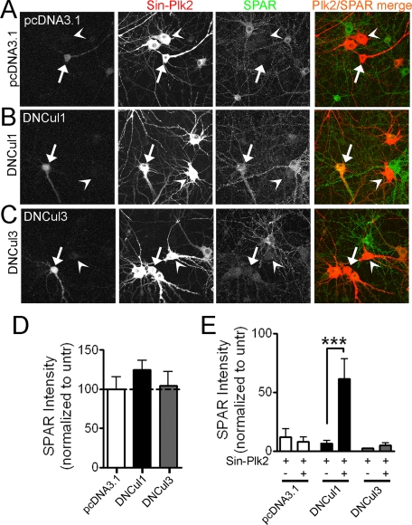
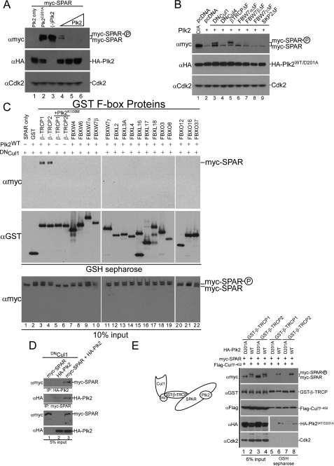
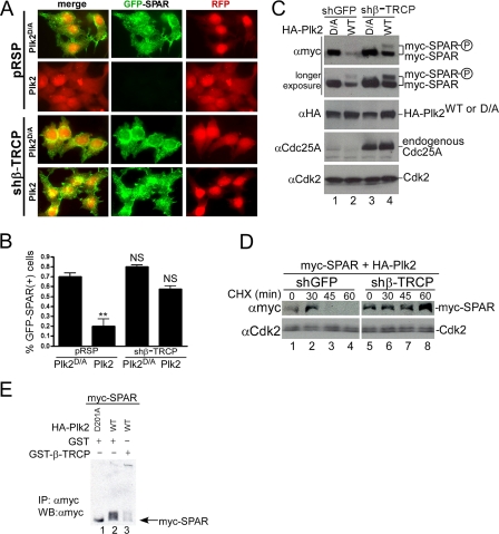
The ubiquitin-proteasome pathway (UPP) regulates synaptic function, but little is known about specific UPP targets and mechanisms in mammalian synapses. We report here that the SCF(beta-TRCP) complex, a multisubunit E3 ubiquitin ligase, targets the postsynaptic spine-associated Rap GTPase activating protein (SPAR) for degradation in neurons. SPAR degradation by SCF(beta-TRCP) depended on the activity-inducible protein kinase Polo-like kinase 2 (Plk2). In the presence of Plk2, SPAR physically associated with the SCF(beta-TRCP) complex through a canonical phosphodegron. In hippocampal neurons, disruption of the SCF(beta-TRCP) complex by overexpression of dominant interfering beta-TRCP or Cul1 constructs prevented Plk2-dependent degradation of SPAR. Our results identify a specific E3 ubiquitin ligase that mediates degradation of a key postsynaptic regulator of synaptic morphology and function.


Figures

FIGURE 1.
FIGURE 1.
Plk2-induced SPAR degradation requires a Cul1-based E3 Ubligase. A-C, dominant negative Cul1 constructs block Plk2-dependent loss of SPAR in hippocampal neurons. Dissociated rat hippocampal neurons (DIV16) were transfected with dominant negative Cullin plasmids (B and C) or a control plasmid (A) and super-infected 2 days later with FLAG-tagged Plk2 driven by Sindbis virus (Sin-Plk2). Neurons were fixed ∼18 h post-infection and immunostained for endogenous SPAR and infected Plk2. Transfected cells were identified during image acquisition by the presence of a co-transfected “fill” protein (GFP, seen in the first column of images). SPAR (green) and Plk2 (red) were pseudo-colored for illustrative purposes after image analysis. Arrows point to cells that are both transfected and infected; arrowheads point to cells that are infected only. Yellow indicates the presence of both SPAR and Plk2 staining. D and E, quantification of SPAR immunostaining in somatic and proximal dendritic regions as integrated immunofluorescence intensity per area in cells transfected with indicated plasmids and/or infected with Plk2 Sindbis virus (Sin-Plk2), normalized to nearby untransfected (untr) cells. Values represent the mean ± S.E., n > 17 cells for all conditions, ***, p < 0.001, Mann-Whitney test (E).
FIGURE 2.
FIGURE 2.
SPAR physically associates with the SCFβ-TRCP complex. A, Plk2-dependent loss of SPAR. HEK293T cells were transfected with 1 μg of pCMV-HA-Plk2 (lane 1), 1 μg of pCMV-myc-SPAR (lane 3), or 1 μg of pCMV-myc-SPAR together with either 1 μg of catalytically inactive pCMV-HA-Plk2D201A (lane 2) or increasing amounts of pCMV-HA-Plk2WT (0.3, 1, or 2 μg) (lanes 4-6). The total amount of transfected DNA was kept constant among all conditions with use of empty vector. Whole cell lysates were immunoblotted with Myc antibody to assess myc-SPAR levels. B, dominant negative versions of Cul1 and β-TRCP stabilize SPAR. pCMV-myc-SPAR (0.5 μg) and pCMV-HA-Plk2D201A/WT (1 μg) (catalytically inactive, lane 1; wild type, lanes 2-9) were co-expressed in HEK293T cells with 2.5 μg of either empty vector, dominant negative Cullins, or dominant negative F-box proteins. Changes in the abundance of myc-SPAR were determined by immunoblotting with anti-Myc antibody. C, F-box protein interaction screen. pCMV-myc-SPAR (0.6 μg), pCMV-HA-Plk2WT/K108M (0.6 μg), and pCMV-DNCul1 (2 μg) were co-expressed as shown with pCMV-GST (lane 2) or the indicated F-box proteins as GST fusions (0.6 μg) (lanes 3-22) in HEK293T cells seeded in 6-well plates. After 24 h, cell extracts were used for GSH-Sepharose pull-down assays, and proteins were immunoblotted with anti-GST and anti-Myc antibodies. Crude lysates were blotted as an input control. D, coimmunoprecipitation of SPAR and Plk2. Extracts of HEK293T cells transfected with pCMV-DNCul1 and pCMV-myc-SPAR (lane 1), pCMV-HA-Plk2 (lane 2), or both (lane 3) were immunoprecipitated using anti-Myc or anti-HA antibodies as indicated. E, formation of a SPAR·Plk2·β-TRCP·Cul1 complex with active Plk2. Lysates from cells transfected with pCMV-myc-SPAR (0.5 μg), pCMV-HA-Plk2WT/D201A (0.5 μg), pCMV-GST-β-TRCP (0.5 μg), and pCMV-FLAG-Cul11-452 (2 μg), as indicated, were incubated with GSH-Sepharose and immunoblotted with anti-Myc, anti-HA, anti-GST, and anti-FLAG antibodies as shown. Lanes 1-4 show lysates (6% of input of the GSH-Sepharose binding reactions).
FIGURE 3.
FIGURE 3.
SCFβ-TRCP regulates Plk2-dependent SPAR abundance, turnover, and promotes its ubiquitination. A and B, depletion of β-TRCP by RNAi protects GFP-SPAR from degradation in individual HEK293T cells. The indicated plasmids were transfected with pCMV-RFP as a co-transfection marker into HEK293T cells. After 96 h, cells were fixed and imaged for GFP-SPAR and RFP expression (A) and quantified as the percentage of RFP expressing cells that also expressed GFP-SPAR (B). Values represent the mean ± S.E. from three independent experiments, derived from analysis of 400 cells per experiment per condition (n = 1200 cells per condition). **, p < 0.01; NS, not significant; one-way analysis of variance, compared with control condition (pRSP+Plk2D/A). C and D, depletion of β-TRCP by RNAi stabilizes SPAR abundance and turnover in the presence of Plk2 activity. HEK293T cells were transfected with vectors expressing myc-SPAR (0.5 μg), WT, or catalytically inactive HA-Plk2 (D/A) (1 μg), and the indicated shRNA vector carrying a puromycin resistance selection marker (2.5 μg). 36 h post-transfection, cells were incubated with media containing 1 μg/ml puromycin to enrich for shRNA expression. Extracts were subsequently examined by immunoblotting 96 h post-transfection as indicated. Endogenous Cdc25A was probed to control for successful knockdown of β-TRCP. In panel D, HEK293T cells were transfected in an identical fashion to panel C, except that 24 h post-transfection the transfected cells were split among 5 wells in puromycin-containing media. After 48 h of puromycin selection, cells were treated with 25 μg/ml cycloheximide (CHX) and harvested at the indicated times before immunoblotting. E, expression of β-TRCP promotes Plk2-mediated SPAR ubiquitination. HEK293T cells were transfected with myc-SPAR (1 μg), His-Ub (1 μg), HA-Plk2 (1.5 μg, wild type or D201A), and GST or GST-β-TRCP (4.5 μg). Twenty hours post-transfection cells were treated with 25 μm MG-132 for 5 h and lysed in buffer containing 10 mm N-ethylmaleimide. Myc-SPAR was purified with c-Myc 9E10-agarose, resolved on 6% Tris-glycine SDS-PAGE gel, and immunoblotted (IB) with anti-Myc antibodies. IP, immunoprecipitates.
FIGURE 4.
FIGURE 4.
A candidateβ-TRCP phosphodegron in SPAR. A, schematic representation of SPAR domains and SPAR fragments: actin-binding domains (Act1 and Act2), RapGAP, PDZ, and guanylate kinase binding (GKBD) domain. The candidate DSGIDT phosphodegron motif (residues 1304-1309) identified in the Act2 domain of SPAR closely resembles the consensus β-TRCP phosphodegron (inset). Boundaries of generated C-terminal fragments of SPAR are depicted; C-1, C-2, and Act2 fragments contain the putative phosphodegron motif, whereas C-3 does not. B and C, SPAR fragments spanning the β-TRCP phosphodegron bind to β-TRCP. HEK293T cells were transfected with vectors expressing SPAR fragments C-1, C-2, C-3 (B) or Act2 (C) (0.6 μg) either alone or with pCMV-GST or pCMV-GST-β-TRCP (0.6 μg). In addition, pCMV-HA-Plk2WT/K108M (0.6 μg) and DNCul1 (2 μg) were co-transfected as indicated. After 24 h, cell extracts were used for GSH-Sepharose pulldown assays, and proteins were immunoblotted with Myc antibodies. Crude lysates were blotted as an input control. D and E, phosphodegron-dependent binding to β-TRCP. Constructs expressing point mutations (S1305A, T1309A) in full-length SPAR (E) and the Act2 fragment (D) (myc-SPARAA or myc-Act2AA, 0.6 μg) were transfected into HEK293T cells along with pCMV-HA-Plk2WT/D201A (0.6 μg), pCMV-DNCul1 (2 μg), and pCMV-GST or pCMV-GST-β-TRCP (0.6 μg) as indicated. Cell lysates were incubated with GSH-Sepharose and immunoblotted with Myc antibodies. Crude extracts were resolved to control for input. Wild type constructs expressing myc-SPAR and myc-Act2 were used for comparison as a positive control for interaction with GST-β-TRCP. F, SPAR associates with endogenous SCFβ-TRCP1 complex in the presence of active Plk2 and dependent upon its phosphodegron. Constructs expressing full-length myc-SPAR (WT or AA) were co-expressed in HEK293T cells with active (WT) or catalytically inactive Plk2 (D201A). Before lysis and immunoprecipitation with 9E10-agarose, cells were treated with proteasome inhibitor MG-132 (25 μm) for 5 h. Proteins bound to 9E10-agarose were analyzed via immunoblotting using Myc antibodies and antibodies that recognized endogenous β-TRCP1 and Cul1. Myc-SPAR-C3, a fragment of SPAR that does not contain the phosphodegron served as a negative control, and myc-Cdc25A, a known target of SCFβ-TRCP1, served as a positive control. G, phosphodegron-dependent degradation of SPAR by Plk2 and β-TRCP. Myc-SPARWT and myc-SPARAA abundance was compared by immunoblotting with the indicated antibodies in HEK293T cells in the absence and presence of pCMV-HA-Plk2 in the background of empty vector or β-TRCPΔF.
FIGURE 5.
FIGURE 5.
Regulation of SPAR degradation in hippocampal neurons through the SCFβ-TRCP complex. A-C, dominant negative β-TRCP constructs block Plk2-dependent loss of SPAR in hippocampal neurons. As in Fig. 1, DIV16 dissociated rat hippocampal neurons were transfected with dominant negative β-TRCP plasmids (A and B) or control SkpΔF plasmid (C) and superinfected 2 days later with FLAG-tagged Plk2 driven by Sindbis virus (Sin-Plk2). Arrows point to cells that are both transfected and infected; arrowheads point to cells that are infected only. Yellow indicates the presence of both SPAR and Plk2 staining. D and E, quantification of SPAR immunostaining in somatic and proximal dendritic regions, as in Fig. 1. Values represent the mean ± S.E. n = 25-37 cells for all constructs in panel D; *, p < 0.05 indicates significant difference from theoretical mean of 100%, Student's t test (D). For panel E, n = 19-37 cells (infected only), n = 13-28 cells (infected and transfected), ***, p < 0.001, Mann Whitney test.

References

    1. Kasai, H., Matsuzaki, M., Noguchi, J., Yasumatsu, N., and Nakahara, H. (2003) Trends Neurosci. 26 360-368 - PubMed
    1. Tada, T., and Sheng, M. (2006) Curr. Opin. Neurobiol. 16 95-101 - PubMed
    1. Bonhoeffer, T., and Yuste, R. (2002) Neuron 35 1019-1027 - PubMed
    1. Carlisle, H. J., and Kennedy, M. B. (2005) Trends Neurosci. 28 182-187 - PubMed
    1. Pak, D. T., Yang, S., Rudolph-Correia, S., Kim, E., and Sheng, M. (2001) Neuron 31 289-303 - PubMed

Publication types

MeSH terms