. 2008 Oct 24;283(43):29196-205.
doi: 10.1074/jbc.M801907200. Epub 2008 Aug 22.

Beta-catenin levels influence rapid mechanical responses in osteoblasts

Affiliations

Beta-catenin levels influence rapid mechanical responses in osteoblasts

Natasha Case et al. J Biol Chem. .

Abstract

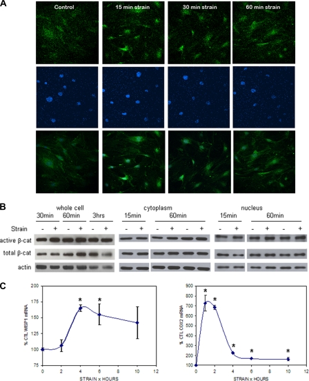
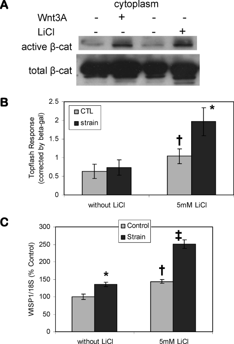
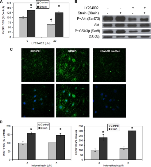
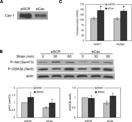
Mechanical loading of bone initiates an anabolic, anticatabolic pattern of response, yet the molecular events involved in mechanical signal transduction are not well understood. Wnt/beta-catenin signaling has been recognized in promoting bone anabolism, and application of strain has been shown to induce beta-catenin activation. In this work, we have used a preosteoblastic cell line to study the effects of dynamic mechanical strain on beta-catenin signaling. We found that mechanical strain caused a rapid, transient accumulation of active beta-catenin in the cytoplasm and its translocation to the nucleus. This was followed by up-regulation of the Wnt/beta-catenin target genes Wisp1 and Cox2, with peak responses at 4 and 1 h of strain, respectively. The increase of beta-catenin was temporally related to the activation of Akt and subsequent inactivation of GSK3beta, and caveolin-1 was not required for these molecular events. Application of Dkk-1, which disrupts canonical Wnt/LRP5 signaling, did not block strain-induced nuclear translocation of beta-catenin or up-regulation of Wisp1 and Cox2 expression. Conditions that increased basal beta-catenin levels, such as lithium chloride treatment or repression of caveolin-1 expression, were shown to enhance the effects of strain. In summary, mechanical strain activates Akt and inactivates GSK3beta to allow beta-catenin translocation, and Wnt signaling through LRP5 is not required for these strain-mediated responses. Thus, beta-catenin serves as both a modulator and effector of mechanical signals in bone cells.


Figures

FIGURE 1.
FIGURE 1.
Mechanical strain induces β-catenin accumulation. A, CIMC-4 cells were subjected to strain for 15–60 min and immunostained for active β-catenin (top) and DAPI (middle). Images of active β-catenin and DAPI staining were merged (bottom) to visualize active β-catenin in the nucleus. Increased nuclear active β-catenin was detected 15 min after beginning mechanical strain by confocal microscopy. B, CIMC-4 cells were strained for 15 min to 3 h. Protein from whole cell lysates and cytoplasmic and nuclear fractionates was extracted and run at 10 μg/lane on 7.5% SDS-PAGE. Western blotting showed increases in active and total β-catenin from whole cell lysates at 30 and 60 min, respectively, whereas an increase in both cytoplasmic and nuclear active β-catenin was detected 60 min after strain. Expression of actin is shown as a control for equal protein loading. C, expression of the β-catenin target genes WISP1 and COX2 in CIMC-4 cells 1–10 h after strain initiation was evaluated by real time RT-PCR. A significant increase in WISP1 expression was measured at 4 and 6 h, whereas COX2 expression was increased at all times. Two experiments grouped (mean ± S.E.) for statistical analyses are shown in the graph. *, significant effect of strain (p < 0.05). CTL, control.
FIGURE 2.
FIGURE 2.
Inhibition of β-catenin proteolysis augments strain effects. A, Western blot for β-catenin in CIMC-4 cells after a 3-h treatment of 20 mm LiCl or 100 ng/ml Wnt3a showed that LiCl increased cytoplasmic active β-catenin to an extent similar to that of Wnt3a. B, CIMC-4 cells were transiently transfected with TopFlash plasmid containing six TCF/LEF binding sites driving the expression of luciferase. β-Galactosidase plasmid was transfected at the same time to control for transfection efficiency. TopFlash response in osteoblasts strained overnight was only seen when cells were also exposed to LiCl (5 mm). Six experiments grouped (mean ± S.E.) for statistical analyses are shown in the graph. *, significant effect of strain (p < 0.05); †, significant difference between unstrained basal and LiCl-treated cultures (p < 0.05). CTL, control. C, strain up-regulation of WISP1 was enhanced in the presence of LiCl. WISP1 expression at 4 h of strain was evaluated by real time RT-PCR. Cultures were treated with LiCl 1 h prior to strain initiation. The data were normalized to the expression level measured in basal control cells. Data were compiled from two experiments and are shown as mean ± S.E. *, significant effect of strain (p < 0.05); †, significant difference between basal and LiCl control cultures (p < 0.05); ‡, significant difference from all other conditions (p < 0.05).
FIGURE 3.
FIGURE 3.
Strain inhibits GSK3β action through an LRP-independent process. A, Western blot of GSK3β phosphorylation (serine 9) showed an increase at 30 min of strain and a peak response at 1 h. Actin protein was blotted as a control for equal protein loading. B, Western blot of Akt phosphorylation (serine 473) showed a strong increase at 15 min of strain, with the response returning to base line at 1 h. C, strain up-regulation of WISP1 and COX2 expression was not disrupted by treatment with the LRP5 inhibitor Dkk-1 (50 ng/ml). WISP1 and COX2 expression at 4 h of strain were evaluated by real time RT-PCR. Cultures were treated with Dkk-1 1 h prior to strain initiation. The data were normalized to the expression level measured in basal control cells. Data were compiled from three experiments and shown as mean ± S.E. *, shows significant effect of strain, p < 0.05. D, Western blots showed that Dkk-1 did not block the increase in phospho-GSK3β (Ser9) and phospho-Akt (Ser473) levels by strain. GSK3β was evaluated at 60 min of strain, and Akt was evaluated at 30 min. E, confocal microscopy showed that Dkk-1 did not block strain-induced nuclear translocation of active β-catenin. Active β-catenin level was assessed by immunofluorescence (top) 15 min after strain initiation. Merged images of active β-catenin and nuclear DAPI staining are shown in the bottom.
FIGURE 4.
FIGURE 4.
Mechanical activation of β-catenin targets does not require PI3-kinase or PGE2 generation. A, WISP1 expression was decreased by treatment with the PI3-kinase inhibitor LY294002 (20 μm), but strain up-regulated WISP1 expression in the presence of LY294002. WISP1 expression at 4 h of strain was evaluated by real-time RT-PCR. Cultures were treated with the inhibitor 1 h prior to strain initiation. The data were normalized to the expression level measured in basal control cells. Data were compiled from two experiments and shown as mean ± S.E. *, significant effect of strain (p < 0.05); †, significant difference between basal and LY294002 control cultures (p < 0.05). B, Western blots showed that strain-induced increases in phospho-GSK3β (PGSK3β) (Ser9), and phospho-Akt (PAkt) (Ser473) were not blocked by LY294002 treatment. C, CIMC-4 cells were subjected to strain for 15 min, and active β-catenin level was assessed by immunofluorescence (top). Merged images of active β-catenin and nuclear DAPI staining are shown in the bottom. Confocal microscopy showed that LY294002 did not block strain-induced nuclear translocation of active β-catenin. A sample in which the active β-catenin antibody was omitted was also included (right). D, strain up-regulation of WISP1 and COX2 expression was not disrupted by treatment with the cyclooxygenase inhibitor indomethacin (5 μm). WISP1 and COX2 expression at 6 h of strain were evaluated by real time RT-PCR. Cultures were treated with indomethacin 1 h prior to strain initiation. The data were normalized to the expression level measured in basal control cells. Representative data are shown as mean ± S.E., and the experiment was repeated one time. *, significant effect of strain (p < 0.05).
FIGURE 5.
FIGURE 5.
Caveolin-1 knockdown does not prevent strain activation of kinase and gene targets. A, caveolin-1 protein levels were decreased by treatment with siRNA targeting caveolin-1 for 48 h. B, Western blot showed that caveolin-1 knockdown did not inhibit strain activation of Akt. Strain-induced inactivation of GSK3β, shown by the increase of phospho-GSK3β (Ser9), was enhanced after strain for 30 min when caveolin-1 expression was decreased. The base-line levels of phospho-Akt (Ser473) and phospho-GSK3β (Ser9) were decreased in cells transfected with siCav. Representative blotting is shown here. Averaged densitometries (mean ± S.E.) for phosphorylated proteins from three experiments are shown in the graphs. C, real time RT-PCR after 4 h of mechanical strain in cells transfected with control siRNA (siSCR) or siCav. The strain effect on WISP1 expression was masked in cells transfected with siSCR. Increases in WISP1 and RUNX2 gene expression by strain were seen in cells treated with siCav. Eight experiments grouped (mean ± S.E.) for statistical analyses are shown in the graph. *, significant effect of strain (p < 0.01).
FIGURE 6.
FIGURE 6.
Caveolin-1 knockdown increases β-catenin nuclear translocation. A, CIMC-4 cells transfected with siRNA targeting caveolin-1 showed increased levels of cytoplasmic active and total β-catenin by Western blot. B, TopFlash response in cells treated with siCav showed increased base-line TopFlash signal. LiCl (20 mm) induced an increase of TopFlash signal, and its effect was further enhanced when caveolin-1 was decreased. Three experiments grouped (mean ± S.E.) for statistical analyses are shown in the graph. **, significant effect of LiCl (p < 0.01); ††, significant difference between control siRNA- and siCav-treated cultures (p < 0.01). C, Western blots of caveolin-1 immunoprecipitates after strain was applied for 15–60 min. The basal association of β-catenin with caveolin-1 was not disrupted by strain application. Caveolin-1 was blotted to show equal immunoprecipitate levels in samples.

References

    1. Rubin, C., and Lanyon, L. (1984) J. Bone Joint Surg. 66A 397-402 - PubMed
    1. Rubin, J., Fan, X., Biskobing, D., Taylor, W., and Rubin, C. (1999) J. Orthop. Res. 17 639-645 - PubMed
    1. Krishnan, V., Bryant, H. U., and Macdougald, O. A. (2006) J. Clin. Invest. 116 1202-1209 - PMC - PubMed
    1. Sawakami, K., Robling, A. G., Ai, M., Pitner, N. D., Liu, D., Warden, S. J., Li, J., Maye, P., Rowe, D. W., Duncan, R. L., Warman, M. L., and Turner, C. H. (2006) J. Biol. Chem. 281 23698-23711 - PubMed
    1. Robinson, J. A., Chatterjee-Kishore, M., Yaworsky, P. J., Cullen, D. M., Zhao, W., Li, C., Kharode, Y., Sauter, L., Babij, P., Brown, E. L., Hill, A. A., Akhter, M. P., Johnson, M. L., Recker, R. R., Komm, B. S., and Bex, F. J. (2006) J. Biol. Chem. 281 31720-31728 - PubMed

Publication types

MeSH terms