. 2008 Oct;7(10):1781-94.
doi: 10.1128/EC.00230-08. Epub 2008 Aug 22.

Copper distributed by Atx1 is available to copper amine oxidase 1 in Schizosaccharomyces pombe

Affiliations

Copper distributed by Atx1 is available to copper amine oxidase 1 in Schizosaccharomyces pombe

Chardeen Peter et al. Eukaryot Cell. 2008 Oct.

Abstract

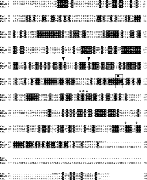
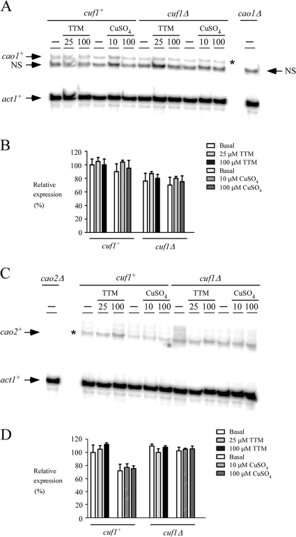
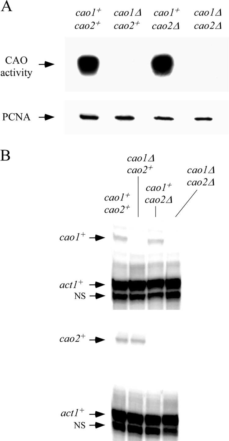
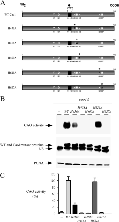
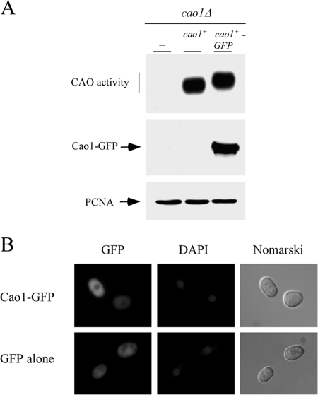
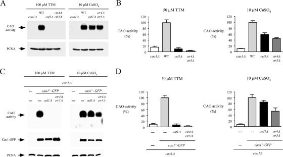
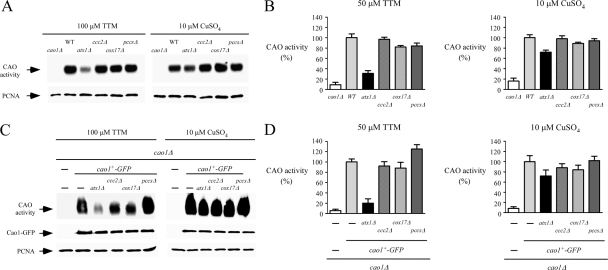
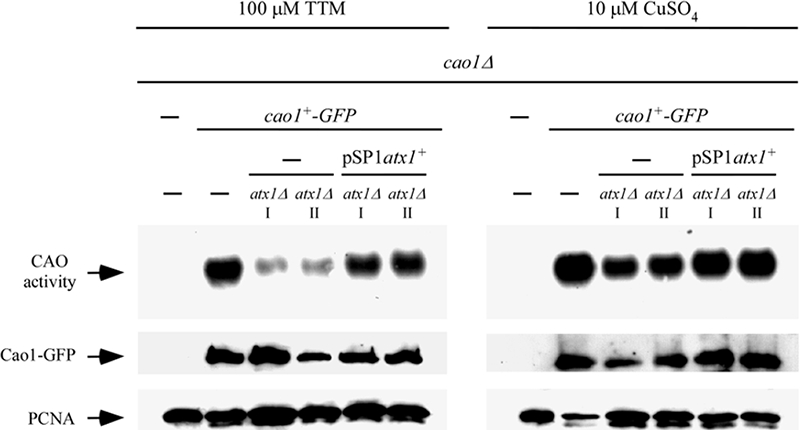
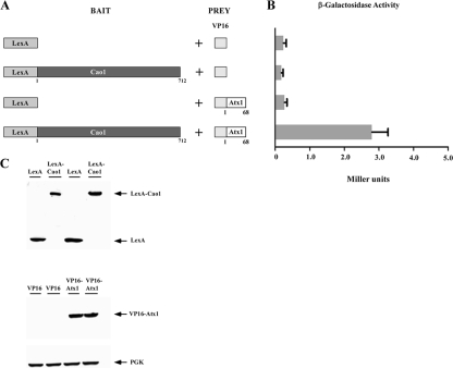
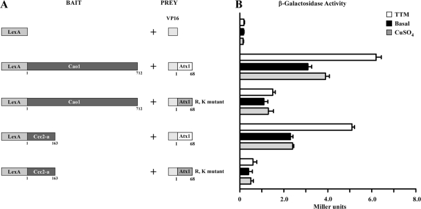
Copper amine oxidases (CAOs) have been proposed to be involved in the metabolism of xenobiotic and biogenic amines. The requirement for copper is absolute for their activity. In the fission yeast Schizosaccharomyces pombe, cao1(+) and cao2(+) genes are predicted to encode members of the CAO family. While both genes are expressed in wild-type cells, we determined that the expression of only cao1(+) but not cao2(+) results in the production of an active enzyme. Site-directed mutagenesis identified three histidine residues within the C-terminal region of Cao1 that are necessary for amine oxidase activity. By use of a cao1(+)-GFP allele that retained wild-type function, Cao1-GFP was localized in the cytosol (GFP is green fluorescent protein). Under copper-limiting conditions, disruption of ctr4(+), ctr5(+), and cuf1(+) produced a defect in amine oxidase activity, indicating that a functionally active Cao1 requires Ctr4/5-mediated copper transport and the transcription factor Cuf1. Likewise, atx1 null cells exhibited substantially decreased levels of amine oxidase activity. In contrast, deletion of ccc2, cox17, and pccs had no significant effect on Cao1 activity. Residual amine oxidase activity in cells lacking atx1(+) can be restored to normal levels by returning an atx1(+) allele, underscoring the critical importance of the presence of Atx1 in cells. Using two-hybrid analysis, we demonstrated that Cao1 physically interacts with Atx1 and that this association is comparable to that of Atx1 with the N-terminal region of Ccc2. Collectively, these results describe the first example of the ability of Atx1 to act as a copper carrier for a molecule other than Ccc2 and its critical role in delivering copper to Cao1.


Figures

FIG. 1.
FIG. 1.
Amino acid alignment of S. pombe Cao1 and Cao2 with H. polymorpha HPAO. Amino acid residues identical in the compared proteins are shown in inverse highlighting. The rectangle shown in the middle portion of the protein sequences indicates the location of the conserved N-Y-E-Y motif in which the first peptidyl tyrosine residue (black circle) is predicted to serve as a precursor for TPQ. Arrowheads indicate putative amino acids that may promote the active conformation of TPQ during the enzymatic reaction. Asterisks show putative histidine residues located in the C-terminal halves of the proteins that are potentially involved in the coordination of one copper atom. The amino acid sequence numbers refer to the position relative to the first amino acid of each protein.
FIG. 2.
FIG. 2.
cao1+ and cao2+ mRNA levels are constitutively expressed and present in cuf1Δ cells. (A) The indicated isogenic strains were grown to logarithmic phase in yeast extract plus supplements. Cultures were incubated in the absence (−) or presence of TTM (25 and 100 μM) or CuSO4 (10 and 100 μM) for 1 h at 30°C. Total RNA was isolated and analyzed by RNase protection assays. Steady-state mRNA levels of cao1+ and act1+ (indicated with arrows) were analyzed in strains expressing (cuf1+) or lacking (cuf1Δ) the cuf1+ allele. As a control, the cao1+ mRNA was not detected in the isogenic cao1Δ null strain. NS, nonspecific signal. The asterisk indicates the cao1+ mRNA. (B) Quantification of results of three independent RNase protection assays, including results shown in panel A. (C) Aliquots of the cultures described for panel A were examined by RNase protection assays for steady-state levels of cao2+ mRNA. The arrows indicate signals corresponding to cao2+ and act1+ steady-state mRNA levels. RNA isolated and analyzed from the isogenic cao2Δ null strain was used as a control. The asterisk indicates the cao2+ mRNA. (D) Quantification of cao2+ mRNA levels after the treatments. The values are the averages from triplicate determinations ± standard deviations.
FIG. 3.
FIG. 3.
Cao1 but not Cao2 catalyzes the oxidative deamination of ethylamine. (A) Four isogenic fission yeast strains (cao1+ cao2+, cao1Δ cao2+, cao1+ cao2Δ, and cao1Δ cao2Δ strains) were analyzed for the presence of CAO activity by use of a chemiluminescence activity assay with ethylamine (10 mM) as a substrate (top). As an internal control, aliquots of total protein extracts were analyzed by immunoblotting using an anti-PCNA antibody (bottom). (B) Total RNA was prepared from aliquots of cultures used in the experiment described for panel A. Representative RNase protection assays of cao1+ (top) and cao2+ (bottom) are shown, indicating steady-state mRNA levels. Actin (act1+) mRNA levels were probed as an internal control. NS, nonspecific signal.
FIG. 4.
FIG. 4.
Functional dissection of critical His residues in Cao1. (A) Schematic representation of the WT Cao1, Cao1 H456A, Cao1 H458A, Cao1 H460A, Cao1 H621A, and Cao1 H627A mutant proteins. The point mutations are marked with asterisks and an A (instead of the WT H residues). The black region indicates the location of the highly conserved consensus sequence NYEY, in which the first peptidyl tyrosine (Y) residue (black circle) serves as a putative precursor for TPQ formation. The active conformation of TPQ is presumably stabilized through interactions with Y307 and D321 in Cao1. The amino acid sequence numbers refer to the position relative to the first amino acid of the protein. (B) CAO activity was determined for cao1Δ cells that were transformed with a plasmid alone (−), WT cao1+, or the indicated mutant alleles of cao1 (top). Protein extracts were prepared from aliquots of cultures and then analyzed by immunoblotting using either anti-His5 or anti-PCNA (as an internal control) antibody. NS, nonspecific signal. (C) Total extracts from cells transformed as described for panel B were assayed for CAO activity using a spectrophotometric method with 4-aminoantipyrine and vanillic acid (23). The CAO activities reported represent the means from three separate determinations ± standard deviations.
FIG. 5.
FIG. 5.
Cytosolic localization of a functional Cao1-GFP fusion protein. (A) S. pombe cells carrying a disrupted cao1Δ allele were transformed with an empty plasmid (−) or a plasmid expressing cao1+ or cao1+-GFP and then tested for the ability to mediate the oxidative deamination of ethylamine using a chemiluminescence activity assay (top). Extracts used to detect CAO activity were also probed with anti-GFP (middle) or anti-PCNA (bottom) antibody. (B) Representative cao1Δ cells expressing Cao1-GFP and GFP alone (as a control) are shown. Cells were grown to exponential phase and then analyzed by direct fluorescence microscopy for GFP. DAPI staining visualized nuclear DNA, and Nomarski optics were used to examine cell morphology.
FIG. 6.
FIG. 6.
Cao1 activity requires expression of the Ctr4/Ctr5 copper transporting complex at the cell surface and the transcription factor Cuf1. (A) The WT and the isogenic cao1Δ, cuf1Δ, and ctr4Δ ctr5Δ mutant strains were incubated in the presence of TTM (100 μM) or CuSO4 (10 μM) and analyzed using an in-gel assay for CAO activity (top). Aliquots of total-extract preparations were assayed by immunoblotting using anti-PCNA antibody (bottom). (B) For a quantitative assay of CAO activity, hydrogen peroxide released from a reaction catalyzed by amine oxidase was determined with a spectrophotometric method using 4-aminoantipyrine and vanillic acid to generate a quinoneimine dye. Each sample was assayed in triplicate. (C) Isogenic strains harboring insertionally inactivated cao1Δ, cuf1Δ cao1Δ, or ctr4Δ ctr5Δ cao1Δ genes were transformed with the GFP-tagged cao1+ allele. After treatment with TTM (100 μM) or CuSO4 (10 μM), whole-cell extracts were prepared and analyzed using an in-gel assay for CAO activity (top). Aliquots of lysates were also analyzed by immunoblotting to verify the presence of Cao1-GFP (middle). Furthermore, all samples were subjected to immunoblotting with anti-PCNA antibody (bottom). As a control, an S. pombe strain bearing a cao1 deletion was transformed with an empty vector (−). (D) Extract preparations from strains described for panel C were analyzed using the spectrophotometric method described for panel B. The reported values of CAO activities are the means from three replicates ± standard deviations.
FIG. 7.
FIG. 7.
Production of fully active Cao1 requires functional Atx1. (A) Logarithmic-phase cultures of isogenic FY435 (WT), cao1Δ, atx1Δ, ccc2Δ, cox17Δ, and pccsΔ strains were treated in the presence of TTM (100 μM) or CuSO4 (10 μM) at 30°C. After 8 h of treatment, cell lysates were prepared from each culture and analyzed using an in-gel peroxidase-catalyzed chemiluminescence assay for CAO activity (top). As an internal control, aliquots of cell lysates were probed by immunoblotting using anti-PCNA antibody (bottom). (B) CAO activity was quantitated from the cell lysates described for panel A by use of a spectrophotometric method with 4-aminoantipyrine and vanillic acid. Error bars indicate the standard deviation of activities from samples analyzed in triplicate. (C) Isogenic S. pombe strains bearing a single deletion (cao1Δ) or a double deletion (atx1Δ cao1Δ, ccc2Δ cao1Δ, cox17Δ cao1Δ, or pccsΔ cao1Δ) were transformed with an integrative plasmid expressing a functional Cao1-GFP fusion protein. Whole-cell extracts from each transformant grown in the presence of TTM (100 μM) or CuSO4 (10 μM) were prepared and analyzed for CAO activity using an in-gel assay (top). Aliquots of total-extract preparations were examined by immunoblotting using either anti-GFP (middle) or anti-PCNA (bottom) antibody. As a control, an S. pombe strain bearing a cao1 deletion was transformed with an empty integrative vector (−). (D) Total-extract preparations from strains described for panel C were analyzed by a spectrophotometric method using 4-aminoantipyrine/vanillic acid. The values of CAO activities are the means from three replicates ± standard deviations.
FIG. 8.
FIG. 8.
Loss of CAO activity in atx1Δ cells is restored by exogenous copper or a plasmid-borne copy of the WT atx1+ gene. An integrative plasmid expressing a functional GFP-tagged cao1+ allele was transformed into cao1Δ and cao1Δ atx1Δ cells. Disruption of the atx1+ gene decreased CAO activity levels. The CAO activity defect was corrected either by returning a WT copy of the atx1+ gene expressed from a plasmid or by adding CuSO4 (10 μM) to the growth medium. Roman numerals (I and II) indicate two separate cao1Δ atx1Δ strains in which cao1+-GFP was returned by integration.
FIG. 9.
FIG. 9.
S. pombe Atx1 interacts with Cao1 by two-hybrid assay. (A) Schematic representation of the LexA DNA binding domain alone or fused upstream of and in frame to the full-length Cao1 coding region. The indicated bait molecule was coexpressed with the VP16 activation domain or the VP16-Atx1 fusion protein. (B) Interactions between the proteins were detected by liquid β-galactosidase assays. The values are the averages from triplicate determinations ± standard deviations. (C) Total-cell-extract preparations from aliquots of cultures used in the assays described for panel B were analyzed by immunoblotting with anti-LexA, anti-VP16, or anti-PGK (as an internal control) antibody.
FIG. 10.
FIG. 10.
The interaction between Cao1 and Atx1 is similar to that of Ccc2-a and Atx1 and is enhanced under low-copper conditions. (A) Schematic diagrams of the LexA DNA binding domain and fusions with Cao1 and the putative N-terminal cytosolic domain of the S. pombe Ccc2 polypeptide (Ccc2-a) are depicted at left. The VP16 and chimeric VP16-Atx1 and VP16-Atx1(R,K) molecules used as prey are shown at right. (B) Mid-logarithmic-phase cells cotransformed with the indicated plasmids were grown under basal or copper-deficient (1 mM TTM) conditions or with excess copper (100 μM CuSO4) for 5 h. Protein-protein interactions were detected by liquid β-galactosidase assays, and results are indicated in Miller units. Error bars indicate the standard deviations of samples analyzed in triplicate.

Similar articles

Cited by

References

    1. Alfa, C., P. Fantes, J. Hyams, M. McLeod, and E. Warbrick. 1993. Experiments with fission yeast, a laboratory course manual. Cold Spring Harbor Laboratory Press, Cold Spring Harbor, NY.
    1. Beaudoin, J., and S. Labbé. 2001. The fission yeast copper-sensing transcription factor Cuf1 regulates the copper transporter gene expression through an Ace1/Amt1-like recognition sequence. J. Biol. Chem. 27615472-15480. - PubMed
    1. Beaudoin, J., and S. Labbé. 2006. Copper induces cytoplasmic retention of fission yeast transcription factor Cuf1. Eukaryot. Cell 5277-292. - PMC - PubMed
    1. Beaudoin, J., J. Laliberté, and S. Labbé. 2006. Functional dissection of Ctr4 and Ctr5 amino-terminal regions reveals motifs with redundant roles in copper transport. Microbiology 152209-222. - PubMed
    1. Beaudoin, J., A. Mercier, R. Langlois, and S. Labbé. 2003. The Schizosaccharomyces pombe Cuf1 is composed of functional modules from two distinct classes of copper metalloregulatory transcription factors. J. Biol. Chem. 27814565-14577. - PubMed

Publication types

MeSH terms

LinkOut - more resources