. 2008 Oct;74(20):6206-15.
doi: 10.1128/AEM.01053-08. Epub 2008 Aug 22.

Identification and characterization of lactococcal-prophage-carried superinfection exclusion genes

Affiliations

Identification and characterization of lactococcal-prophage-carried superinfection exclusion genes

Jennifer Mahony et al. Appl Environ Microbiol. 2008 Oct.

Abstract

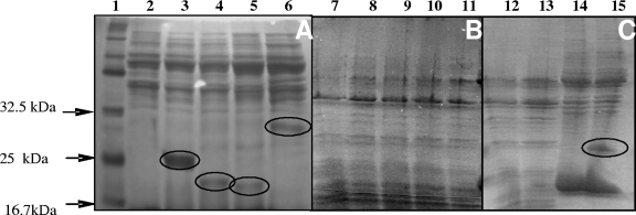
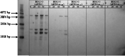
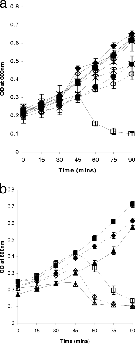
Superinfection exclusion (Sie) proteins are prophage-encoded phage resistance systems. In this study, genes encoding Sie systems were identified on the genomes of Lactococcus lactis subsp. cremoris MG1363 and L. lactis subsp. lactis IL1403. These Sie systems are genetically distinct and yet were shown to act specifically against a particular subset of the 936 phage group. Each of the systems allows normal phage adsorption while affecting plasmid transduction and intracellular phage DNA replication, which points to the blocking of phage DNA injection as their common mode of action. Sie-specifying genes found on the MG1363 prophages are also present in various lactococcal strains, whereas the prophage-encoded Sie systems of IL1403 do not appear to be as widely disseminated.


Figures

FIG. 1.
FIG. 1.
Gradient polyacrylamide gel (10 to 20%) of the membrane vesicle (A) and cytoplasmic fractions (B) of L. lactis NZ9000(pNZ8048), NZ9000(pNZ8048-sie309), NZ9000(pNZ8048-sie312), NZ9000(pNZ8048-siemg2), and NZ9000(pNZ8048-sieT712). Lane 1, protein molecular weight marker; lanes 2 to 6, membrane vesicle fractions of the strains listed in the above order; lanes 7 to 11, cytoplasmic fractions of the strains listed in the above order. Each of the expressed Sie proteins is circled in lanes 3 to 6. Panel C represents SDS-PAGE analysis of the cytoplasmic and membrane vesicle fractions of L. lactis NZ9000(pNZ8048) and NZ9000(pNZ8048-sie2009), with the band representing Sie2009 circled in the membrane vesicle fraction in lane 15. Lanes 12 and 13, cytoplasmic fractions of L. lactis NZ9000(pNZ8048) and NZ9000(pNZ8048-sie2009), respectively. Lanes 14 and 15, membrane vesicle fractions of L. lactis NZ9000(pNZ8048) and NZ9000(pNZ8048-sie2009), respectively.
FIG. 2.
FIG. 2.
Southern blot analysis of sk1 intracellular DNA replication. Total cellular DNA isolated at intervals of 20 min after infection over a total period of 60 min, as indicated in the picture. Lanes 1 to 4, MG1363(pNZ44) infected with sk1; lanes 5 to 8, MG1363(pNZ44sie309) infected with sk1; lanes 9 to 12, MG1363(pNZ44sie312) infected with sk1; lanes 13 to 16, MG1363(pNZ44siemg2) infected with sk1; lanes 17 to 20, MG1363(pNZ44sieT712) infected with sk1. DNA was digested with Sau3AI and blotted and probed with the PCR-amplified orf18 gene of sk1.
FIG. 3.
FIG. 3.
(a) Lysis in broth experiments comparing the lysis profiles of MG1363 harboring pNZ44 in both uninfected cells (▪) and cells infected (□) with sk1 and its derivatives bearing siemg2 (uninfected, X with short dashes; infected, X with long dashes), sieT712 (uninfected, ▴; infected ▵), sie309 (uninfected, •; infected, ○, and sie312 (uninfected, ⧫; infected ⋄). OD600 readings were taken at 15-min intervals. Each of the active Sie proteins provides resistance against sk1 infection. MG1363 harboring pNZ44 was used as a positive control. Each assay was performed in triplicate. (b) Lysis in broth experiments of L. lactis MG1363(pNZ44) cells both uninfected (⧫) and infected (⋄) with sk1 and its derivatives bearing the putative Sie genes orf2141mg3 (uninfected, ⧫; infected, ⋄) and orf2286 (uninfected, ▪; infected, □). OD600 readings were taken at 15-min intervals. These systems appear to provide little or no protection against infection by sk1.

Similar articles

Cited by

References

    1. Ammann, A., H. Neve, A. Geis, and K. J. Heller. 2008. Plasmid transfer via transduction from Streptococcus thermophilus to Lactococcus lactis. J. Bacteriol. 190:3083-3087. - PMC - PubMed
    1. Brondsted, L., S. Ostergaard, M. Pedersen, K. Hammer, and F. K. Vogensen. 2001. Analysis of the complete DNA sequence of the temperate bacteriophage TP901-1: evolution, structure, and genome organization of lactococcal bacteriophages. Virology 283:93-109. - PubMed
    1. Burgess, C., M. O'Connell-Motherway, W. Sybesma, J. Hugenholtz, and D. van Sinderen. 2004. Riboflavin production in Lactococcus lactis: potential for in situ production of vitamin-enriched foods. Appl. Environ. Microbiol. 70:5769-5777. - PMC - PubMed
    1. Chandry, P. S., S. C. Moore, J. D. Boyce, B. E. Davidson, and A. J. Hillier. 1997. Analysis of the DNA sequence, gene expression, origin of replication and modular structure of the Lactococcus lactis lytic bacteriophage sk1. Mol. Microbiol. 26:49-64. - PubMed
    1. Chibani-Chennoufi, S., M.-L. Dillmann, L. Marvin-Guy, S. Rami-Shojaei, and H. Brussow. 2004. Lactobacillus plantarum bacteriophage LP65: a new member of the SPO1-like genus of the family Myoviridae. J. Bacteriol. 186:7069-7083. - PMC - PubMed

Publication types