. 2008 Sep 2;105(35):13027-32.
doi: 10.1073/pnas.0805038105. Epub 2008 Aug 22.

Dysregulation of microRNAs after myocardial infarction reveals a role of miR-29 in cardiac fibrosis

Affiliations

Dysregulation of microRNAs after myocardial infarction reveals a role of miR-29 in cardiac fibrosis

Eva van Rooij et al. Proc Natl Acad Sci U S A. .

Abstract

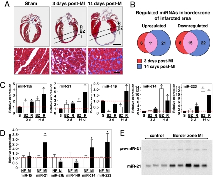
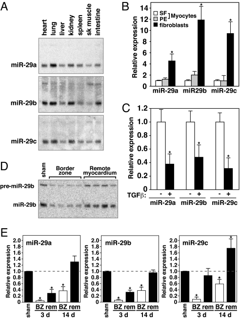
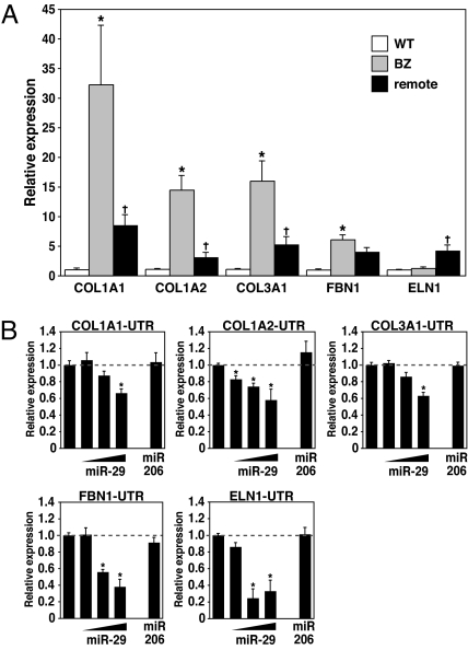
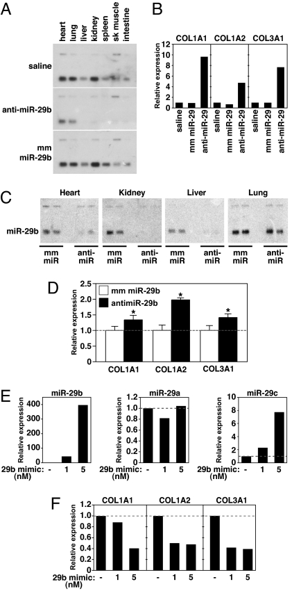
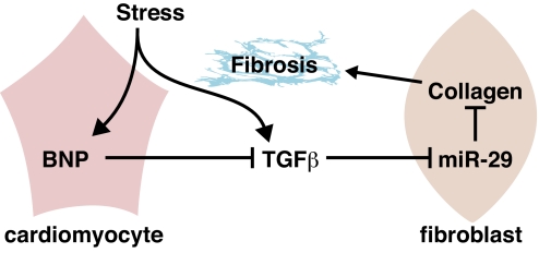
Acute myocardial infarction (MI) due to coronary artery occlusion is accompanied by a pathological remodeling response that includes hypertrophic cardiac growth and fibrosis, which impair cardiac contractility. Previously, we showed that cardiac hypertrophy and heart failure are accompanied by characteristic changes in the expression of a collection of specific microRNAs (miRNAs), which act as negative regulators of gene expression. Here, we show that MI in mice and humans also results in the dysregulation of specific miRNAs, which are similar to but distinct from those involved in hypertrophy and heart failure. Among the MI-regulated miRNAs are members of the miR-29 family, which are down-regulated in the region of the heart adjacent to the infarct. The miR-29 family targets a cadre of mRNAs that encode proteins involved in fibrosis, including multiple collagens, fibrillins, and elastin. Thus, down-regulation of miR-29 would be predicted to derepress the expression of these mRNAs and enhance the fibrotic response. Indeed, down-regulation of miR-29 with anti-miRs in vitro and in vivo induces the expression of collagens, whereas over-expression of miR-29 in fibroblasts reduces collagen expression. We conclude that miR-29 acts as a regulator of cardiac fibrosis and represents a potential therapeutic target for tissue fibrosis in general.


Conflict of interest statement

Conflict of interest statement: E.v.R., W.S.M., and E.N.O. are cofounders of MiRagen Therapeutics.

Figures

Fig. 1.
Fig. 1.
MiRNA profiling in response to MI. (A) Masson Trichrome staining of mouse heart sections shows early scar formation 3 days after MI, with myocyte hypertrophy, loss of myocytes, and collagen deposition. Fourteen days after MI, there is a thin stretched infarct that results in cardiac hypertrophy and interstitial fibrosis in the border zone of the infarcted region. I, infarct; BZ, borderzone; R, remote myocardium. (Lower) Higher magnification of the borderzone regions of the infarcted hearts and the comparable level of the sham operated heart. (Scale bars: Upper, 2 mm; Lower, 20 μm). (B) Microarray analysis reveals miRNAs are dynamically regulated in response to MI. Even after initial infarct healing, 14 days after MI, 11 miRs are overlappingly up-regulated, whereas 15 miRs are down-regulated. The number of miRNAs regulated ≥2-fold in each category is shown. (C) Real-time PCR analysis confirms the regulation of specific miRNAs in response to MI compared with sham operated animals (n = 3–4. S, sham; BZ, borderzone; R, remote. *, P < 0.05 compared with sham operated animals). (D) Real-time PCR analysis shows the regulation of miRNAs in human heart samples in response to MI compared with nonfailing hearts (n = 5–6, NF = nonfailing, MI = myocardial infarction). *, P < 0.05 compared with nonfailing hearts. (E) Northern blot analysis of three nonfailing human hearts and five human hearts after MI indicates a consistent increase in miR-21 in the borderzone of human heart samples in response to MI.
Fig. 2.
Fig. 2.
Down-regulation of miR-29 in the infarcted region after MI. (A) Northern blot analysis of mouse tissues indicates a large overlap in expression of all three miR-29 members, with highest expression in lung, heart, and kidney. Of the miR-29 members, miR-29b appeared most highly expressed in the heart. (B) Real-time PCR analysis indicated all three members of the miR-29 family to be highly expressed in fibroblasts compared with cardiomyocytes either under serum free conditions (SF) or stimulated with phenylephrine (PE). Comparable amounts of RNA were used in each reaction. *, P < 0.05 compared with untreated myocytes. (C) Real-time PCR analysis indicates that all three miR-29 family members are down-regulated in fibroblasts after exposure to TGFβ for 48 h. *, P < 0.05 compared with untreated fibroblasts. (D) Northern blot analysis on mouse cardiac tissue 3 days after MI shows a consistent down-regulation of miR-29 in response to MI compared with sham-operated animals. Down-regulation is more pronounced in the borderzone than in the remote myocardium. The Northern blot shows the level of miR-29b in four different animals both in the borderzone and the corresponding remote area. (E) Real-time analysis indicates all miR-29 members to be regulated in response to MI. Whereas the down-regulation is most pronounced in the border zone (BZ) of the infarct 3 days after MI, this down-regulation remains present even after initial infarct healing has taken place. (n = 3–4 per group. BZ, borderzone; rem, remote. *, P < 0.05 compared with sham operated animals.)
Fig. 3.
Fig. 3.
miR-29 regulates extracellular matrix protein mRNAs. (A) Real-time PCR analysis of predicted target genes in both the borderzone and remote myocardium 3 days after MI shows a decrease in miR-29 to correlate with an increase in collagens (COL1A1, COL1A2, and COL3A1) and fibrillin (FBN1), whereas there was no significant change in elastin (ELN1). (n = 3–4 per group. BZ, borderzone. *, P < 0.05 compared with sham operated animals, + P < 0.05 compared with BZ region). (B) COS cells were transiently transfected with luciferase reporters (100 μg) linked to the 3′UTR sequences of the indicated genes. Increasing amounts of the miR-29b-1/miR-29a cluster (25–50-100 μg) repress the expression of luciferase, whereas this decrease was absent when using an unrelated miR, miR-206 (50 μg). *, P < 0.05 compared with luciferase reporter alone.
Fig. 4.
Fig. 4.
miR-29 inhibition induces fibrosis in vivo. (A) Northern blot analysis showing knockdown of miR-29b expression three days after i.v. injection of 80 mg/kg of either anti-miR-29 or mm miR-29 or a comparable volume of saline. (B) Real-time PCR analysis of liver extracts reveals a pronounced increase in collagen expression in response to miR-29 knockdown, whereas this effect was absent after saline or mm injection. (C) Northern blot of the indicated tissues 3 weeks after i.v. injection with 80 mg/kg on two consecutive days of either anti-miR-29 or mm miR-29 oligonucleotide or a comparable volume of saline. miR-29 is nearly abolished in the liver, heart and kidney, whereas miR-29 levels in lung appear unaffected by anti-miR-29. (D) Real-time PCR analysis of heart extracts indicates an increase in cardiac collagen expression in response to miR-29 knockdown. (n = 2 per group, *, P < 0.05 compared with mm treated animals). (E) Cardiac fibroblasts were either left untreated or treated with 1 or 5 nM miR-29b mimic for 48 h. Real-time PCR analysis indicates an increase in miR-29b expression in fibroblasts two days after miR-29b mimic treatment, whereas miR-29a levels were unchanged and miR-29c levels only slightly increased. (F) Cardiac fibroblasts that were either left untreated or treated with 1 or 5 nM miR-29b mimic for 48 h show a decrease in expression of collagen genes in response to increased levels of miR-29b as determined by real-time PCR analysis.
Fig. 5.
Fig. 5.
A model for the role of miR-29 in cardiac fibrosis. In response to cardiac stress, TGFβ is activated and triggers the down-regulation of miR-29 in cardiac fibroblasts and consequent up-regulation of the expression of collagens and other ECM proteins involved in fibrosis. At the same time, stress induces the expression of anti-fibrotic BNP, which is secreted by cardiomyocytes and counteracts the activation and thereby profibrotic function of TGFβ.

References

    1. Fox CS, et al. Increasing cardiovascular disease burden due to diabetes mellitus: The Framingham Heart Study. Circulation. 2007;115:1544–1550. - PubMed
    1. Swynghedauw B. Molecular mechanisms of myocardial remodeling. Physiol Rev. 1999;79:215–262. - PubMed
    1. Bouzeghrane F, Reinhardt DP, Reudelhuber TL, Thibault G. Enhanced expression of fibrillin-1, a constituent of the myocardial extracellular matrix in fibrosis. Am J Physiol Heart Circ Physiol. 2005;289:H982–991. - PubMed
    1. Powell DW, et al. Myofibroblasts. I. Paracrine cells important in health and disease. Am J Physiol. 1999;277:C1–C9. - PubMed
    1. Cheng Y, et al. MicroRNAs are aberrantly expressed in hypertrophic heart: Do they play a role in cardiac hypertrophy? Am J Pathol. 2007;170:1831–1840. - PMC - PubMed

Publication types

MeSH terms