. 2008 Aug 26;105(34):12581-6.
doi: 10.1073/pnas.0803174105. Epub 2008 Aug 22.

Lack of adrenomedullin in the mouse brain results in behavioral changes, anxiety, and lower survival under stress conditions

Affiliations

Lack of adrenomedullin in the mouse brain results in behavioral changes, anxiety, and lower survival under stress conditions

Ana P Fernández et al. Proc Natl Acad Sci U S A. .

Abstract

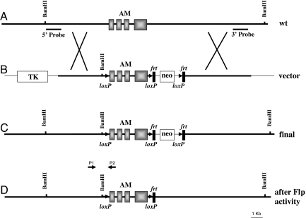
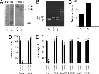
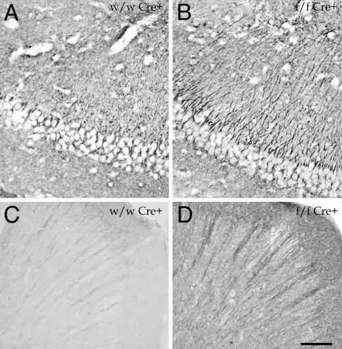
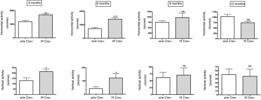
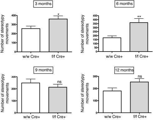
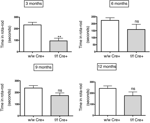
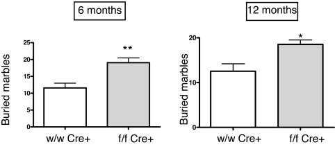
The adrenomedullin (AM) gene, adm, is widely expressed in the central nervous system (CNS) and several functions have been suggested for brain AM. Until now, a formal confirmation of these actions using genetic models has been elusive since the systemic adm knockout results in embryo lethality. We have built a conditional knockout mouse model using the Cre/loxP approach. When crossed with transgenic mice expressing the Cre recombinase under the tubulin Talpha-1 promoter, we obtained animals with no AM expression in the CNS but normal levels in other organs. These animals lead normal lives and do not present any gross morphological defect. Specific areas of the brain of animals lacking CNS AM contain hyperpolymerized tubulin, a consequence of AM downregulation. Behavioral analysis shows that mice with no AM in their brain have impaired motor coordination and are hyperactive and overanxious when compared to their wild-type littermates. Treatment with methylphenidate, haloperidol, and diazepam did not show differences between genotypes. Circulating levels of adrenocorticotropic hormone and corticosterone were similar in knockout and wild-type mice. Animals with no brain AM were less resistant to hypobaric hypoxia than wild-type mice, demonstrating the neuroprotective function of AM in the CNS. In conclusion, AM exerts a beneficial action in the brain by maintaining homeostasis both under normal and stress conditions.


Conflict of interest statement

The authors declare no conflict of interest.

Figures

Fig. 1.
Fig. 1.
Gene targeting of adm. (A) Structure of the wild-type allele as found in mouse chromosome 7. The 4 exons of the adm gene are represented as solid boxes. Southern probes at the 5′ and 3′ ends of the construct are also indicated. (B) Targeting vector designed to insert loxP sequences (arrowheads) around the adm gene and the neomycin cassette (neo) surrounded by frt sequences (vertical rectangles). An additional BamHI site was added next to the first loxP to allow for identification. The TK gene was also added as a negative marker. A thinner line represents vector sequences. (C) Structure of the modified allele in the ES cells after recombination takes place. (D) Structure of the final allele, following the crossing with Flp expressing mice. P1 (AMD sense) and P2 (AMD antisense) are the primers for diagnostic PCR described in Table S1.
Fig. 2.
Fig. 2.
Characterization of the mice lacking brain AM. (A) Southern blot analysis of BamHI-digested ES cell DNA with the external 5′ probe (Left) and the internal 3′ probe (Right). The wild-type (w) allele renders a band of 17 kb whereas the mutant alleles (f) render 4- and 12-kb bands, respectively. (B) PCR analysis of tail DNA from 3-week-old mice with AMD primers (Table S1). After PCR, samples were digested with BamHI. The wild-type (w) allele produces a band of ≈400 bp whereas the mutant (f) is identified by a 200-bp band. Heterozygous mice (w/f) produce both bands. (C) Genotypes of offspring issued by the cross of 5 pairs of heterozygous mice for the ubiquitous deletion. No homozygous mutant mice (−/−) were produced. (D) Real-time PCR quantification of the levels of AM produced in the brain and heart of wild type (wt/wt Cre+) and brain conditional AM mutants (f/f Cre+). While a large diminution is appreciated in the mutant brain, the heart AM contents do not show any significant difference with the wild type. (E) Real-time PCR quantification of the levels of AM and AM-related molecules CLR, RAMPs, AM2, and CGRP in the brain of wild-type and mutant animals. The drastic reduction of AM in the mutant brain does not affect the expression of the related genes.
Fig. 3.
Fig. 3.
Immunohistochemical staining for Glu-tubulin in the hippocampus (A and B) and frontal cortex (C and D) of wild type (A and C) and brain conditional AM mutants (B and D). Mutant animals show a higher staining intensity than normal mice. (Scale bar: A and B, 20 μm; C and D, 100 μm.)
Fig. 4.
Fig. 4.
Activity cage measurements of horizontal (Upper) and vertical (Lower) activity in male mice at different ages. Mutant animals lacking brain AM (gray bars) are more active than their wild-type littermates (open bars) during the first 6 months of life. Bars represent the mean ± standard deviation of 10 mice. The whole experiment was repeated twice, and a representative example is presented here. *, P < 0.05; **, P < 0.01; ***, P < 0.001; ns, no significant differences.
Fig. 5.
Fig. 5.
Stereotypy quantification as recorded in the activity cage in male mice at different ages. Mutant animals (f/f Cre+) present a higher number of stereotypies than wild-type littermates (wt/wt Cre+) for the first 6 months but the difference disappears in older mice. Bars represent the mean ± standard deviation of 10 mice. The whole experiment was repeated 3 times, and a representative example is presented here. *, P < 0.05; **, P < 0.01; ns, no significant differences.
Fig. 6.
Fig. 6.
Rotarod measurements in male mice at different ages, expressed as the time in seconds the animal stayed in the rod. Mutant animals lacking brain AM (gray bars) are less coordinated than their wild-type littermates (open bars) at 3 months. Bars represent the mean ± standard deviation of 10 mice and 5 trials per animal. The whole experiment was repeated twice, and a representative example is presented here. **, P < 0.01; ns, no significant differences.
Fig. 7.
Fig. 7.
Anxiety assay based on the number of glass marbles buried by the mice in a 30 min period. Mutant animals lacking brain AM (gray bars) are more anxious than their wild-type littermates (open bars). Bars represent the mean ± standard deviation of 10 mice. The whole experiment was repeated twice, and a representative example is presented here. *, P < 0.05; **, P < 0.01.

References

    1. Lopez J, Martinez A. Cell and molecular biology of the multifunctional peptide, adrenomedullin. Int Rev Cytol. 2002;221:1–92. - PubMed
    1. McLatchie LM, et al. RAMPs regulate the transport and ligand specificity of the calcitonin-receptor-like receptor. Nature. 1998;393:333–339. - PubMed
    1. Ueta Y, et al. Adrenomedullin-immunoreactive neurons in the paraventricular and supraoptic nuclei of the rat. Neurosci Lett. 1995;202:37–40. - PubMed
    1. Serrano J, et al. Distribution of adrenomedullin-like immunoreactivity in the rat central nervous system by light and electron microscopy. Brain Res. 2000;853:245–268. - PubMed
    1. Ueda T, Ugawa S, Saishin Y, Shimada S. Expression of receptor-activity modifying protein (RAMP) mRNAs in the mouse brain. Brain Res Mol Brain Res. 2001;93:36–45. - PubMed

Publication types