. 2008 Sep 2;105(35):12903-8.
doi: 10.1073/pnas.0803001105. Epub 2008 Aug 22.

RPA phosphorylation facilitates mitotic exit in response to mitotic DNA damage

Affiliations

RPA phosphorylation facilitates mitotic exit in response to mitotic DNA damage

Rachel William Anantha et al. Proc Natl Acad Sci U S A. .

Abstract

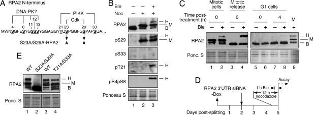
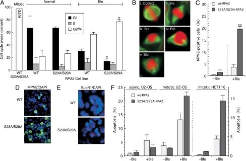
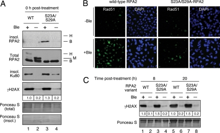
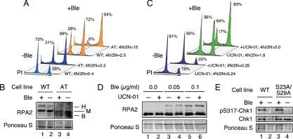
Human replication protein A (RPA) becomes phosphorylated on the RPA2 subunit by cyclin B-Cdc2 during mitosis, although the functional role of this modification is unclear. We find that this modification stimulates RPA2 to become hyperphosphorylated in response to mitotic DNA damage caused by bleomycin treatment. Cells in which endogenous RPA2 was replaced by a mutant subunit lacking both Cdc2 sites had a significant defect in mitotic release into a 2N G(1) phase after exposure to bleomycin. An increased percentage of these mutant cells also was positive initially for cyclin B expression and BubR1 chromatin staining, indicative of an extended spindle assembly checkpoint. The mutant cells that experienced mitotic DNA damage also underwent apoptosis at higher levels than cells expressing the WT subunit. Even so, we did not find the mutation had any dramatic effects on the level of DNA repair in mitosis. Cells lacking ATM (a checkpoint factor and RPA2 kinase) also were severely defective in mitotic exit and were unable to support RPA hyperphosphorylation after mitotic DNA damage. Although checkpoint 1 effector kinase (Chk1) had a more complex role, inhibition of Chk1 activity with UCN-01 also reduced mitotic exit. Chk1 activation and mitotic RPA hyperphosphorylation were found to be independent events. Our results demonstrate that mitotic RPA hyperphosphorylation facilitates release of cells from a damaged mitosis into a 2N G(1) phase, thereby increasing cell viability.


Conflict of interest statement

The authors declare no conflict of interest.

Figures

Fig. 1.
Fig. 1.
Mitotic phosphorylation of RPA2 cyclin-Cdk sites stimulates subsequent stress-dependent modification. (A) Schematic of RPA2 phosphorylation sites. (B) Lysates were isolated from asynchronous nonstressed cells (lane 1) or from nocodazole-arrested cells isolated by mitotic shake-off and then were mock treated (lane 2) or were treated with Ble (2 μg/ml) for 1 h (lane 3). (C) Nocodazole-arrested cells, either treated with Ble (2 μg/ml) for 1 h or not treated, were released into medium lacking both agents and were collected 6 h after release. To examine G1 cells, mitotic cells were released from the nocodazole block and 3 h after release were treated with Ble (2 μg/ml) for 1 h or were mock treated. Cells were collected immediately (0 h) or 4 h after the medium was replaced with medium lacking Ble. (D) Schematic indicating steps involved in cellular RPA2 replacement. −Dox = removal of doxycycline causing the induction of ectopic RPA2 expression. (E) Endogenous RPA2 was silenced in U2-OS cells replacing S23A/S29A-, T21A/S33A-, or WT-RPA2. After silencing (48 h), cells were treated with nocodazole for 12 h. The mitotic cells were collected by shake-off and were treated with Ble (2 μg/ml) for 1 h, followed by collection of cells. For all samples, lysates were prepared from the collected cells and analyzed by Western blotting using the indicated antibodies. The RPA2 species are indicated as follows: B, basal; H, hyperphosphorylated; M, mitotic.
Fig. 2.
Fig. 2.
Loss of mitotic RPA2 phosphorylation inhibits mitotic exit and stimulates cellular apoptosis. (A) Endogenous RPA2 was silenced in U2-OS cells ectopically expressing WT- or S23A/S29A-RPA2. After silencing (48 h), cells were treated with nocodazole for 12 h; then mitotic cells were collected by mechanical shake-off. Cells were mock treated or treated with Ble (2 μg/ml Ble for 1 h), followed by release into medium lacking nocodazole and Ble. Cells were collected 7 h after release and were analyzed by FACS for DNA content. (B) After mitotic release (2 h), cells expressing S23A/S29A- or WT-RPA2 were stained with α-tubulin (to visualize spindles) and 7-amino-actinomycin D (to stain DNA). i shows untreated cells undergoing metaphase. Panels ii through v show Ble-treated cells displaying abnormal spindle assembly and globular DNA appearance. (C and D) Cells were silenced for endogenous RPA2 and ectopic RPA2 was induced. After silencing (48 h), cells were treated with nocodazole for 12 h and were mock treated or treated with Ble (2 μg/ml for 1 h). Then the medium was replaced with medium lacking nocodazole and Ble for 3 h. Cells then were stained and imaged for MPM2 (green) and DAPI (blue) and were quantitated. (E) Mitotic cells expressing ectopic RPA2 and silenced for endogenous RPA2 were mock treated or treated with Ble (2 μg/ml for 1 h) and, 3 h after release from nocodazole and Ble, were stained for BubR1 (red) and DAPI (blue). (F) Asynchronous U2-OS clones were mock treated or treated with Ble (30 μg/ml Ble for 2.5 h); then the medium was replaced with fresh medium. Cells then were analyzed for apoptosis by a TUNEL assay 20 h after release. Mitotically arrested cells were treated with 10 μg/ml Ble for 1 h and then were similarly treated. An identical treatment was used for HCT116 clones, except that cells were scored 8 h after release.
Fig. 3.
Fig. 3.
DNA damage and repair factors following mitotic DNA damage. (A) Mitotic cells expressing WT- or S23A/S29A-RPA2 were mock treated or treated with Ble (2 μg/ml for 1 h) in the presence of nocodazole, followed by extraction of the soluble fraction with CSK buffer containing 0.5% Triton X-100. The insoluble and total fractions then were subjected to Western blot analysis using indicated antibodies. (B) Mitotic cells, as described in A, were released from nocodazole and Ble treatment and were fixed 8 h after release. Cells were then analyzed for Rad51 chromatin association by immunofluorescence microscopy. (C) Mitotic cells as described in A were released into fresh media lacking nocodazole and Ble for either 8 or 20 h and then were isolated. Samples were analyzed for γH2AX levels by Western blot.
Fig. 4.
Fig. 4.
Chk1 and ATM enhance release into G1 on release from genotoxic stress during mitosis. (A) Mitotic WT (GM637) or AT cells (GM5849) were mock treated or treated with Ble (0.05 μg/ml for 1 h). Cells were released into medium lacking nocodazole and Ble and, 3 h after release, were analyzed for DNA content by FACS. (B) Treatment of cells was performed as described in A. Immediately after treatment, samples were collected and analyzed by Western blot for RPA2. (C) Nocodazole-arrested U2-OS cells were mock treated or treated with Ble (0.05 μg/ml for 1 h) in the presence or absence of 300 nM UCN-01. Cells then were released into medium lacking Ble but containing UCN-01 for 3 h and were analyzed by FACS for DNA content. (D) Samples prepared as described in C were treated with either 0.05 or 0.1 μg/ml Ble and were analyzed for RPA2 (and RPA2 hyperphosphorylation) by Western blot. (E) Cells replaced for WT- or S23A/S29A-RPA2 were mock treated or treated with Ble (10 μg/ml for 1 h). Lysates were prepared and analyzed for total Chk1 and pS317-Chk1 by Western blot. Similar results were observed using 2 μg/ml Ble (data not shown). B, basal (nonphosphorylated); H, hyperphosphorylated; M, mitotic.

Similar articles

Cited by

References

    1. Sanchez Y, et al. Control of the DNA damage checkpoint by chk1 and rad53 protein kinases through distinct mechanisms. Science. 1999;286:1166–1171. - PubMed
    1. Skoufias DA, Lacroix FB, Andreassen PR, Wilson L, Margolis RL. Inhibition of DNA decatenation, but not DNA damage, arrests cells at metaphase. Mol Cell. 2004;15:977–990. - PubMed
    1. Iftode C, Daniely Y, Borowiec JA. Replication protein A (RPA): The eukaryotic SSB. Crit Rev Biochem Mol Biol. 1999;34:141–180. - PubMed
    1. Binz SK, Sheehan AM, Wold MS. Replication protein A phosphorylation and the cellular response to DNA damage. DNA Repair (Amst) 2004;3:1015–1024. - PubMed
    1. Zou L, Elledge SJ. Sensing DNA damage through ATRIP recognition of RPA-ssDNA complexes. Science. 2003;300:1542–1548. - PubMed

Publication types