. 2008 Aug 26;105(34):12182-7.
doi: 10.1073/pnas.0804700105. Epub 2008 Aug 22.

Androgen receptor is a tumor suppressor and proliferator in prostate cancer

Affiliations

Androgen receptor is a tumor suppressor and proliferator in prostate cancer

Yuanjie Niu et al. Proc Natl Acad Sci U S A. .

Abstract

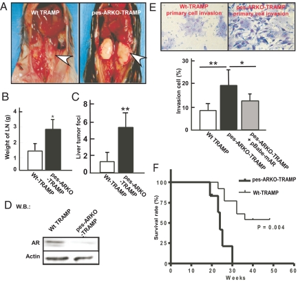
Targeting androgens/androgen receptor (AR) functions via androgen deprivation therapy (ADT) remains the standard treatment for prostate cancer. However, most tumors eventually recur despite ADT. Here we demonstrate that the prostate AR may function as both a suppressor and a proliferator to suppress or promote prostate cancer metastasis. Results from orthotopically recombining stromal WPMY1 cells with epithelial PC3 prostate cancer cells in mice demonstrated that restoring AR in epithelial PC3 cells or knockdown of AR in stromal WPMY1 cells suppressed prostate cancer metastasis. Knockdown of the AR in epithelial CWR22rv1 prostate cancer cells also resulted in increased cell invasion in vitro and in vivo. Restoring AR in PC3 cells (PC3-AR9) results in decreased invasion in bone lesion assays and in vivo mouse models. Mice lacking the prostate epithelial AR have increased apoptosis in epithelial luminal cells and increased proliferation in epithelial basal cells. The consequences of these two contrasting results led to the expansion of CK5/CK8-positive intermediate cells, and mice developed larger and more invasive metastatic tumors in lymph nodes and died earlier than wild-type littermates. Mechanistic dissection suggested that androgens/AR might directly or indirectly modulate metastasis-related genes and suppression of TGFbeta1 signals results in the partial inhibition of AR-mediated metastasis. Collectively, our understanding of these opposing roles of prostatic AR may revolutionize the way we combat prostate cancer, and allow the development of new and better therapies by targeting only the proliferative role of AR.


Conflict of interest statement

The authors declare no conflict of interest.

Figures

Fig. 1.
Fig. 1.
Epithelial AR suppresses and stromal AR stimulates prostate cancer cell invasion. (A) AR protein expression in PC3 cells transfected with AR cDNA under the control of a natural proximal AR promoter region (PC3-AR7, PC3-AR8, and PC3-AR9), strong SV40 promoter (PC3-AR2), or vector only (PC3-v). (B) 1 nM DHT treatment increased AR transactivation of (ARE)4-Luc activity in PC3-AR9 cells. (C) The invasion of PC3-AR9 was decreased as compared to PC3-v with 1 nM DHT using Matrigel-coated Boyden chambers. (D–F) Two-chamber cell recombination assays showed stromal AR stimulated, whereas epithelial AR suppressed, prostate cancer cell invasion. (D) PC3-v or PC3-AR9 cells on the upper layer of the Boyden chamber were cocultured with WPMY1-v or WPMY1-ARsi cells on the lower layer of the chamber. (E) Invasion of PC3-v or PC3-AR9 cells was significantly higher when cocultured with WPMY1-v cells than when cultured with WPMY1-ARsi cells. (F) The quantitative data from experiments (n = 3) *, P < 0.05, **, P < 0.01. (G) Genetic knockdown of AR resulted in increased invasion ability in human prostate cancer CWR22rv1 cells. The alleles of the AR gene in CWR22rv1-AR+/+ cells were genetically disrupted by homologous recombination strategy to produce CWR22rv1-AR+/− cells. Western blot analysis indicated that expression of AR is knocked down in the presence or absence of 1 nM DHT in CWR22rv1-AR+/− cells compared with CWR22rv1-AR+/+ cells (Upper). AR transactivation is diminished in CWR22rv1-AR+/− compared with CWR22rv1-AR+/+ in the presence of 1 nM DHT (Lower Left). The cell invasion (n = 5) increased in CWR22rv1-AR+/− compared with CWR22rv1-AR+/+ in the Boyden chamber assay (Lower Right). *P < 0.05 between the two cell lines. (H) knockdown of AR with siRNA in CWR22rv1-AR+/+cells increased cell invasion, and knock-in of the AR in CWR22rv1-AR+/− cells decreased cell invasion using Boyden chamber assays (n = 5). * Indicates a significant difference between the two transfectants with P < 0.05.
Fig. 2.
Fig. 2.
Adding functional AR to PC3 cells (PC3-AR9) decreased prostate cancer cell invasion in bone lesion test and in preclinical animal models. (A) PC3-AR9 cells formed fewer osteolytic lesions than PC3-v cells. Osteoclasts and osteoclast precursors (OC) were cultured with PC3-v and PC3-AR9 cells on cortical bone wafers. After 10 days, the bone wafers were scraped, dried, and stained with the OC cell indicator tartrate-resistant acid phosphatase (TRAP). The extent of bone resorption with PC3-AR9 cells decreased compared to PC3-v cells as shown by measurement of the area of osteoclast lacunae on the bone wafers. OC alone and OC with parathyroid hormone (PTH) were used as the negative and positive control, respectively. Data were from three independent experiments and are presented as mean ± SD *P < 0.05, **P < 0.01. (B, C) PC3-AR9 cells had less bone invasion compared to PC3-v cells. Effects of intratibial injection of PC3-v and PC3-AR9 cells in nude mice. PC3-v cells developed larger osteolytic lesions than PC3-AR9 cells at 6–8 wks (B, representative x-ray radiograph shown), and larger and more invasive tumors as measured by dial caliper at week 12 (C, arrows and quantitated tumor volume, Lower). Data are shown as mean ± SD; *, P < 0.05; **, P < 0.01. (D) PC3-AR9 cells generated smaller metastatic PLN tumors compared to those generated by PC3-v cells. PC3-v and PC3-AR9 cells (5 × 10⁵) suspended in 100 μl Matrigel were inoculated into the anterior prostate of 16-wk-old nude mice. Twelve weeks after injection, large PC3 prostate tumors developed (not shown). To visualize PLN-metastatic tumors, we removed prostates containing primary tumors. Note that larger PLN-metastatic tumors (shown by gross and H&E staining, Left) were developed in mice inoculated with PC3-v tumors (arrows). The tumor volume was quantitated (right panel, n = 5). (E) PC3-AR9 cells recombined with WPMY1-v or WPMY1-ARsi cells generated smaller metastatic tumors than their control PC3-v recombined groups (arrows). PC3-v or PC3-AR9 cells (5 × 10⁵), combined with WPMY1-v or WPMY1-ARsi cells, were suspended in 100 μl of Matrigel and inoculated into the anterior prostate of 16-wk-old nude mice. Twelve weeks after injection, the tumors developed (not shown), and the sizes of PLN metastatic tumors were compared via gross appearance and histology (H&E). The tumor volume was quantitated (Right, n = 5).
Fig. 3.
Fig. 3.
Loss of epithelial AR in pes-ARKO-TRAMP leads to higher proliferation in basal cells and higher apoptosis in luminal cells with increased basal intermediate cells. (A) Using the TUNEL assay, the apoptosis signals in the luminal epithelial cells from 16-wk-old pes-ARKO-TRAMP prostatic epithelium were higher than those from Wt-TRAMP (arrowheads). CK8 immunostaining (red) was used to identify the luminal epithelial cells (Upper). Quantitative results showed the differences were 18% vs. 2% (Lower). (B) The growth rates of prostate epithelium were demonstrated by Ki67 immunostaining (Top) and BrdU incorporation (Middle) in 16-wk-old Wt-TRAMP vs. pes-ARKO-TRAMP. The mice were i.p. injected with BrdU (10 μg/g body weight) every 6 h, and killed 24 h later. Paraffin-fixed tissue sections were stained by the BrdU detecting kit (Zymed Laboratories). The results from double immunofluorescent staining of BrdU (green) and CK5 (red) indicated that the higher proliferative prostate cells belong to the CK5 positive-basal cells in 16 wk-old pes-ARKO-TRAMP (Bottom with overlapped image, yellow color). The quantitative data were shown (n = 3). (C) Loss of the AR in the epithelium of pes-ARKO-TRAMP led to the expansion of the basal intermediate cell populations (yellow), with increased CK5/CK8-positive signals in prostates of 16-wk-old pes-ARKO-TRAMP compared to Wt-TRAMP mice (Upper). The quantitative data are shown (Lower, n = 3). (D) Increased CD44-positive cells in primary tumors of 24-wk-old pes-ARKO-TRAMP compared to Wt-TRAMP littermates (n = 3).
Fig. 4.
Fig. 4.
pes-ARKO-TRAMP mice develop more aggressive and invasive tumors than Wt-TRAMP littermates. (A) PLN metastases are significantly larger in 24-wk-old pes-ARKO-TRAMP mice compared to Wt-TRAMP mice. (B) Weights of PLN metastases isolated from 24-wk-old pes-ARKO-TRAMP were greater than those of Wt-TRAMP mice (n = 7 mice in each group). (C) The number of liver tumor foci was increased in pes-ARKO-TRAMP mice compared to Wt-TRAMP mice (n = 6 mice in each group). (D) Western blot analysis of AR protein in PLN tumor, from 24-wk-old Wt-TRAMP or pes-ARKO-TRAMP mice. (E) Higher invasiveness of primary cultured PLN tumor cells from pes-ARKO-TRAMP mice as compared to those from Wt-TRAMP mice using Boyden chamber invasion assays (Upper). Adding functional AR by retrovirus infection into primary-cultured pes-ARKO-TRAMP tumor cells results in suppression of invasion (Lower). The purity and originality of primary cultured PLN tumor cells was confirmed by the expression of pan-CK epithelial cell marker (data not shown). Data were presented as mean ± SD (n = 5); *, P < 0.05; **, P < 0.01. (F) survival rate was decreased in pes-ARKO-TRAMP (C57BL/6 × TRAMP-FVB, n = 10) as compared with Wt-TRAMP (C57BL/6 x TRAMP-FVB, n = 16).

References

    1. Chang CS, Kokontis J, Liao ST. Molecular cloning of human and rat complementary DNA encoding androgen receptors. Science. 1988;240:324–326. - PubMed
    1. Heinlein CA, Chang C. Androgen receptor in prostate cancer. Endocr Rev. 2004;25:276–308. - PubMed
    1. Miyamoto H, Messing EM, Chang C. Does androgen deprivation improve treatment outcomes in patients with low-risk and intermediate-risk prostate cancer? Nat Clin Pract Oncol. 2005;2:236–237. - PubMed
    1. Fluchter SH, Weiser R, Gamper C. The role of hormonal treatment in prostate cancer. Recent Results Cancer Res. 2007;175:211–237. - PubMed
    1. Mizokami A, Yeh SY, Chang C. Identification of 3′,5′-cyclic adenosine monophosphate response element and other cis-acting elements in the human androgen receptor gene promoter. Mol Endocrinol. 1994;8:77–88. - PubMed

Publication types