. 2008 Nov 15;112(10):4298-307.
doi: 10.1182/blood-2008-05-156000. Epub 2008 Aug 22.

Targeted deletion of alpha-adducin results in absent beta- and gamma-adducin, compensated hemolytic anemia, and lethal hydrocephalus in mice

Affiliations

Targeted deletion of alpha-adducin results in absent beta- and gamma-adducin, compensated hemolytic anemia, and lethal hydrocephalus in mice

Raymond F Robledo et al. Blood. .

Abstract

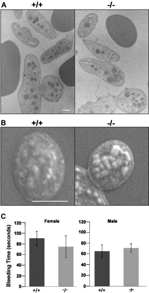
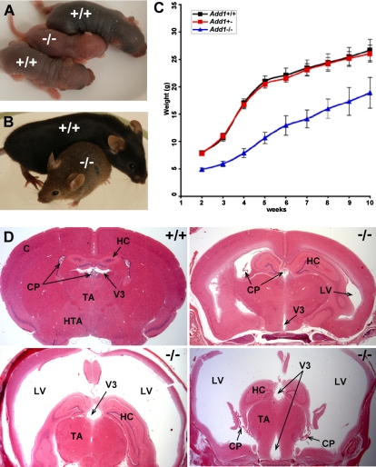
In the red blood cell (RBC), adducin is present primarily as tetramers of alpha- and beta-subunits at spectrin-actin junctions, or junctional complexes. Mouse RBCs also contain small amounts of gamma-adducin. Platelets contain alpha- and gamma-adducin only. Adducin functions as a barbed-end actin capping protein to regulate actin filament length and recruits spectrin to the ends of actin filaments. To further define adducin's role in vivo, we generated alpha-adducin knockout mice. alpha-Adducin is absent in all tissues examined in homozygous null mice. In RBCs, beta- and gamma-adducin are also absent, indicating that alpha-adducin is the limiting subunit in tetramer formation at the spectrin-actin junction. Similarly, gamma-adducin is absent in alpha-null platelets. alpha-Adducin-null mice display compensated hemolytic anemia with features characteristic of RBCs in hereditary spherocytosis (HS), including spherocytes with significant loss of surface area, decreased mean corpuscular volume (MCV), cell dehydration, and increased osmotic fragility. Platelets maintain their normal discoid shape, and bleeding times are normal. alpha-Adducin-null mice show growth retardation at birth and throughout adulthood. Approximately 50% develop lethal communicating hydrocephalus with striking dilation of the lateral, third, and fourth ventricles. These data indicate that adducin plays a role in RBC membrane stability and in cerebrospinal fluid homeostasis.


Figures

Figure 1
Figure 1
Targeting of the α-adducin gene, Add1. (A) Structure of the α-adducin locus according to Ensembl transcript identification ENSMUST00000114340 with the targeted allele shown below. Numbered boxes represent exons. Relevant restriction sites are indicated. (B) Homologous recombination generates a 13.1-kb wild-type and a 9.0-kb mutant allele upon digestion of ES cell DNA with EcoRI. (C) Northern blot analysis of E14.5 fetal liver with hybridization probes indicated. RT-PCR primers used to generate probes are given in Table 1. +/+ indicates wild- type; +/−, heterozygotes; and −/−, homozygous null.
Figure 2
Figure 2
Detection of gene products. (A) Western blots of RBC ghost proteins demonstrating loss of both β- and γ-adducin in α-adducin knockout mice. (B) Northern blots of E14.5 fetal livers showing loss of α-adducin mRNA but normal levels of β- and γ-adducin mRNA. The exon 10 to 12 α-adducin probe was used (Table 1). (C) Western blots demonstrating loss of α-adducin in all tissues examined. (D) Western blots demonstrating loss of platelet γ-adducin in α-adducin–null mice. +/+ indicates wild-type; +/−, heterozygotes; and −/−, homozygous null.
Figure 3
Figure 3
Membrane skeleton in α-adducin–null RBCs. The major membrane skeleton components α- and β-spectrin, ankyrin, band 3, proteins 4.1 and 4.2, and actin appear normal in α-adducin–null RBC ghost membranes by SDS-PAGE (A) and Western blotting (B). Other components of the junctional complex—p55, GPC, and protein 4.9 (dematin)—also appear normal. (C) The actin capping protein EcapZ is strikingly up-regulated in adducin-null RBC membranes. Tropomodulin (Tmod) appears normal. (D) TM is decreased to approximately 20% of normal in the absence of adducin. +/+ indicates wild-type; −/−, homozygous null.
Figure 4
Figure 4
Adducin-null RBCs show features of HS. (A) RBC volume and hemoglobin concentration histograms indicate a population of small (left-shifted volume histogram) and dehydrated (right-shifted hemoglobin concentration histogram) in α-adducin–null (−/−, bottom) versus wild-type (+/+, top) RBCs. (B) Peripheral blood smears reveal a significant population of spherocytes (formula image
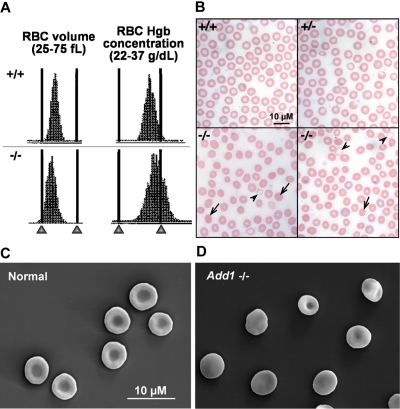
) with a small population of severely hypochromic cells (formula image). +/+ indicates wild-type; +/−, heterozygotes; and −/−, homozygous null. Bar represents 10 μM. (C) Biconcave RBCs from a normal littermate and (D) an α-adducin–null mouse showing significant spherocytosis. Bar represents 10 μM.
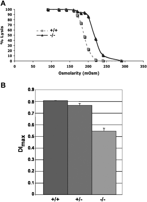
Figure 5
Figure 5
Increased osmotic fragility and ektacytometry. (A) Osmotic fragility curves indicate increased osmotic fragility in α-adducin–null RBCs. (B) The maximum value of the deformability index, DImax, at 250 dynes/cm2 is a direct measure of the mean RBC membrane surface area and is significantly reduced (P < .001) in α-adducin–null RBCs, confirming loss of membrane surface area (X ± SE). +/+ indicates wild-type; +/−, heterozygotes; and −/−, homozygous null.
Figure 6
Figure 6
Platelet structure and function. TEM (A) and SEM (B) reveal adducin-null platelets maintain their normal discoid state in the circulation. (C) Bleeding times. X plus or minus SE.
Figure 7
Figure 7
Growth retardation and lethal hydrocephalus in α-adducin–null mice. (A-C) α-Adducin–null mice are smaller at birth and throughout life compared with their normal littermates. Blue in panel C indicates X plus or minus SE. (D) Approximately 50% of α-adducin–null mice develop hydrocephalus with extreme dilation of the lateral and third ventricles. The fourth ventricle is also enlarged and the cerebral aqueduct is open (not shown), indicating communicating hydrocephalus. Significant thinning of the cortex is apparent as hydrocephalus worsens. Hydrocephalus was not observed in wild-type or heterozygous mice. C indicates cerebral cortex; CP, choroid plexus; HC, hippocampus; TA, thalamus; HTA, hypothalamus; LV, lateral ventricle; V3, third ventricle; +/+, wild-type; and −/−, homozygous null.

References

    1. Suriyapperuma SP, Lozovatsky L, Ciciotte SL, Peters LL, Gilligan DM. The mouse adducin gene family: alternative splicing and chromosomal localization. Mammalian Genome. 2000;11:16–23. - PubMed
    1. Bennett V, Baines AJ. Spectrin and ankyrin-based pathways: Metazoan inventions for integrating cells into tissues. Physiol Rev. 2001;81:1353–1392. - PubMed
    1. Gilligan DM, Lozovatsky L, Gwynn B, Brugnara C, Mohandas N, Peters LL. Targeted disruption of the beta adducin gene (Add2) causes red blood cell spherocytosis in mice. Proc Natl Acad Sci U S A. 1999;96:10717–10722. - PMC - PubMed
    1. Hughes CA, Bennett V. Adducin: a physical model with implications for function in assembly of spectrin-actin complexes. J Biol Chem. 1995;270:18990–18996. - PubMed
    1. Gilligan DM, Bennett V. The junctional complex of the membrane skeleton. Semin Hematol. 1993;30:74–83. - PubMed

Publication types

MeSH terms