

Embryonic stem cells attenuate myocardial dysfunction and inflammation after surgical global ischemia via paracrine actions
- PMID: 18723770
- PMCID: PMC2593500
- DOI: 10.1152/ajpheart.00236.2008
Embryonic stem cells attenuate myocardial dysfunction and inflammation after surgical global ischemia via paracrine actions
Abstract
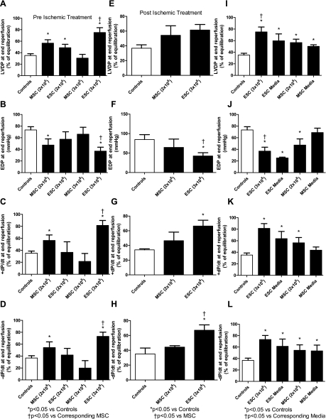
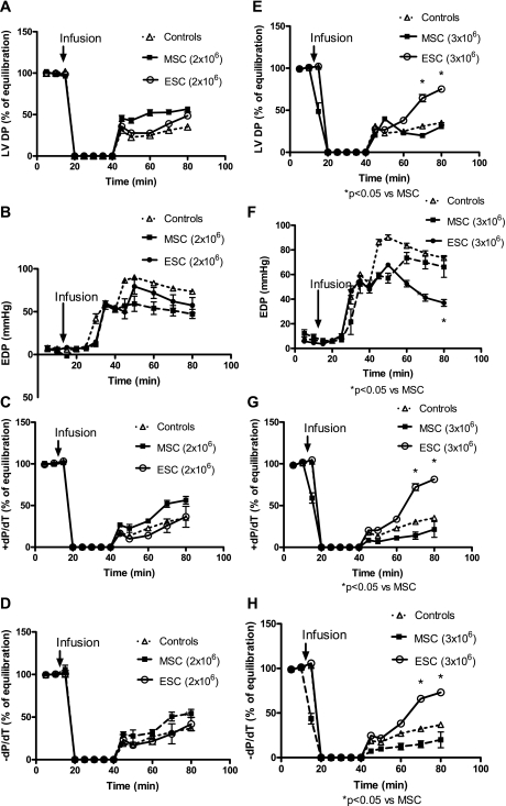
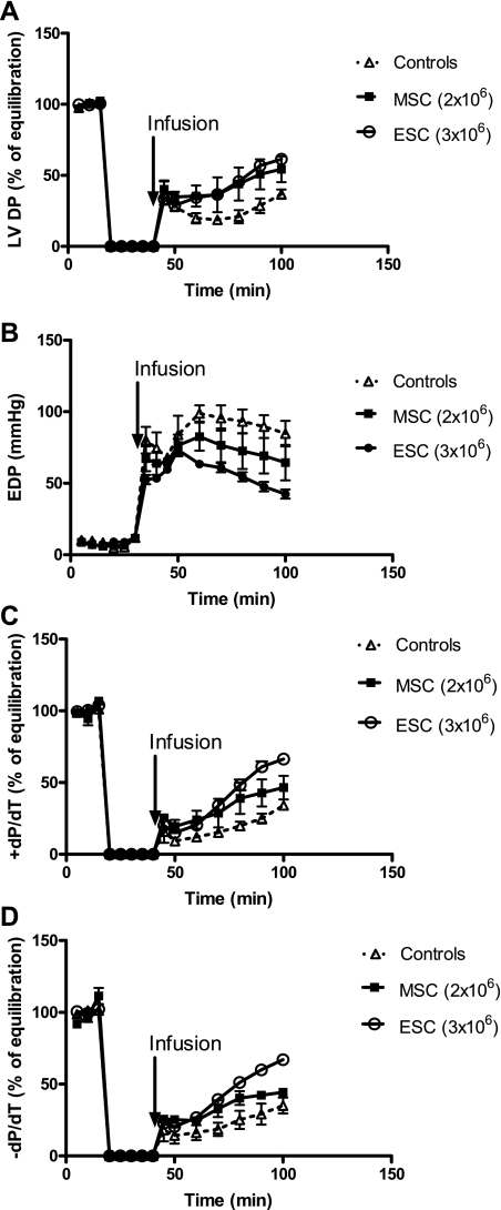
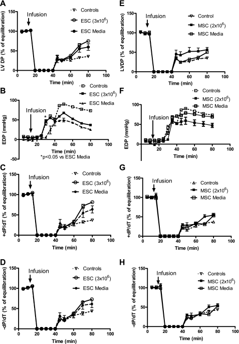
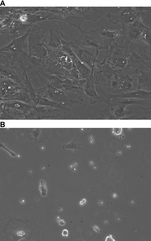
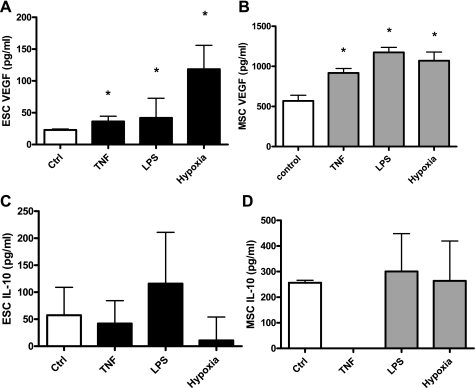
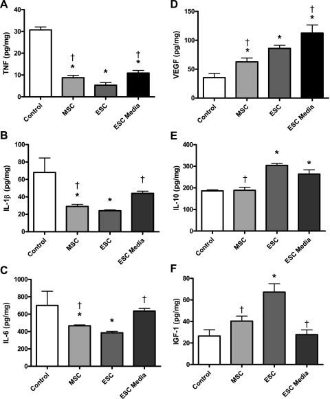
Stem cell treatment may positively influence recovery and inflammation after shock by multiple mechanisms, including the paracrine release of protective growth factors. Embryonic stem cells (ESCs) are understudied and may have greater protective power than adult bone marrow stem cells (BMSCs). We hypothesized that ESC paracrine protective mechanisms in the heart (decreased injury by enhanced growth factor-mediated reduction of proinflammatory cytokines) would be superior to the paracrine protective mechanisms of the adult stem cell population in a model of surgically induced global ischemia. Adult Sprague-Dawley rat hearts were isolated and perfused via Langendorff model. Hearts were subjected to 25 min of warm global ischemia and 40 min of reperfusion and were randomly assigned into one of four groups: 1) vehicle treated; 2) BMSC or ESC preischemic treatment; 3) BMSC or ESC postischemic treatment; and 4) BMSC- or ESC-conditioned media treatment. Myocardial function was recorded, and hearts were analyzed for expression of tissue cytokines and growth factors (ELISA). Additionally, ESCs and BMSCs in culture were assessed for growth factor production (ELISA). ESC-treated hearts demonstrated significantly greater postischemic recovery of function (left ventricular developed pressure, end-diastolic pressure, and maximal positive and negative values of the first derivative of pressure) than BMSC-treated hearts or controls at end reperfusion. ESC-conditioned media (without cells) also conferred cardioprotection at end reperfusion. ESC-infused hearts demonstrated increased VEGF and IL-10 production compared with BMSC hearts. ESC hearts also exhibited decreased proinflammatory cytokine expression compared with MSC hearts. Moreover, ESCs in cell culture demonstrated greater pluripotency than MSCs. ESC paracrine protective mechanisms in surgical ischemia are superior to those of adult stem cells.
Figures
Similar articles
-
VEGF is critical for stem cell-mediated cardioprotection and a crucial paracrine factor for defining the age threshold in adult and neonatal stem cell function.Am J Physiol Heart Circ Physiol. 2008 Dec;295(6):H2308-14. doi: 10.1152/ajpheart.00565.2008. Epub 2008 Oct 10. Am J Physiol Heart Circ Physiol. 2008. PMID: 18849336 Free PMC article.
-
Estradiol-treated mesenchymal stem cells improve myocardial recovery after ischemia.J Surg Res. 2009 Apr;152(2):319-24. doi: 10.1016/j.jss.2008.02.006. Epub 2008 Mar 13. J Surg Res. 2009. PMID: 18511080 Free PMC article.
-
Estradiol treatment promotes cardiac stem cell (CSC)-derived growth factors, thus improving CSC-mediated cardioprotection after acute ischemia/reperfusion.Surgery. 2014 Aug;156(2):243-52. doi: 10.1016/j.surg.2014.04.002. Epub 2014 Jun 21. Surgery. 2014. PMID: 24957669
-
Gene regulatory networks in embryonic stem cells and brain development.Birth Defects Res C Embryo Today. 2009 Jun;87(2):182-91. doi: 10.1002/bdrc.20149. Birth Defects Res C Embryo Today. 2009. PMID: 19530135 Free PMC article. Review.
-
Regenerative Cardiovascular Therapies: Stem Cells and Beyond.Int J Mol Sci. 2019 Mar 21;20(6):1420. doi: 10.3390/ijms20061420. Int J Mol Sci. 2019. PMID: 30901815 Free PMC article. Review.
Cited by
-
High glucose concentration in cell culture medium does not acutely affect human mesenchymal stem cell growth factor production or proliferation.Am J Physiol Regul Integr Comp Physiol. 2009 Jun;296(6):R1735-43. doi: 10.1152/ajpregu.90876.2008. Epub 2009 Apr 22. Am J Physiol Regul Integr Comp Physiol. 2009. PMID: 19386985 Free PMC article.
-
Apelin-13 increases myocardial progenitor cells and improves repair postmyocardial infarction.Am J Physiol Heart Circ Physiol. 2012 Sep 1;303(5):H605-18. doi: 10.1152/ajpheart.00366.2012. Epub 2012 Jun 29. Am J Physiol Heart Circ Physiol. 2012. PMID: 22752632 Free PMC article.
-
Embryonic stem cells for severe heart failure: why and how?J Cardiovasc Transl Res. 2012 Oct;5(5):555-65. doi: 10.1007/s12265-012-9356-9. Epub 2012 Mar 13. J Cardiovasc Transl Res. 2012. PMID: 22411322 Review.
-
Postinfarct intramyocardial injection of mesenchymal stem cells pretreated with TGF-alpha improves acute myocardial function.Am J Physiol Regul Integr Comp Physiol. 2010 Jul;299(1):R371-8. doi: 10.1152/ajpregu.00084.2010. Epub 2010 May 19. Am J Physiol Regul Integr Comp Physiol. 2010. PMID: 20484699 Free PMC article.
-
Gender dimorphisms in progenitor and stem cell function in cardiovascular disease.J Cardiovasc Transl Res. 2010 Apr;3(2):103-13. doi: 10.1007/s12265-009-9149-y. J Cardiovasc Transl Res. 2010. PMID: 20376198 Free PMC article. Review.
References
-
- Ayala A, Herdon CD, Lehman DL, Ayala CA, Chaudry IH. Differential induction of apoptosis in lymphoid tissues during sepsis: variation in onset, frequency, and the nature of the mediators. Blood 87: 4261–4275, 1996. - PubMed
-
- Ayala A, Xin Xu Y, Ayala CA, Sonefeld DE, Karr SM, Evans TA, Chaudry IH. Increased mucosal B-lymphocyte apoptosis during polymicrobial sepsis is a Fas ligand but not an endotoxin-mediated process. Blood 91: 1362–1372, 1998. - PubMed
-
- Balsam LB, Wagers AJ, Christensen JL, Kofidis T, Weissman IL, Robbins RC. Haematopoietic stem cells adopt mature haematopoietic fates in ischaemic myocardium. Nature 428: 668–673, 2004. - PubMed
-
- Crisostomo PR, Markel TA, Wang M, Lahm T, Lillemoe KD, Meldrum DR. In the adult mesenchymal stem cell population, source gender is a biologically relevant aspect of protective power. Surgery 142: 215–221, 2007. - PubMed
Publication types
MeSH terms
Substances
Grants and funding
LinkOut - more resources
Full Text Sources
Other Literature Sources