. 2008 Oct;295(4):H1657-68.
doi: 10.1152/ajpheart.00141.2008. Epub 2008 Aug 22.

Ether-linked diglycerides inhibit vascular smooth muscle cell growth via decreased MAPK and PI3K/Akt signaling

Affiliations

Ether-linked diglycerides inhibit vascular smooth muscle cell growth via decreased MAPK and PI3K/Akt signaling

Kristy L Houck et al. Am J Physiol Heart Circ Physiol. 2008 Oct.

Abstract

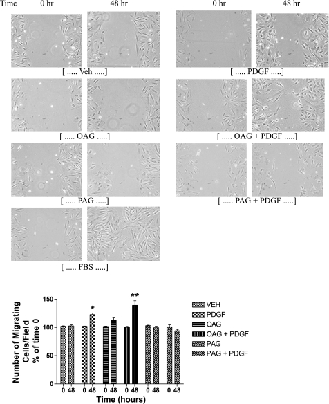
Diglycerides (DGs) are phospholipid-derived second messengers that regulate PKC-dependent signaling pathways. Distinct species of DGs are generated from inflammatory cytokines and growth factors. Growth factors increase diacyl- but not ether-linked DG species, whereas inflammatory cytokines predominately generate alkyl, acyl- and alkenyl, acyl-linked DG species in rat mesenchymal cells. These DG species have been shown to differentially regulate protein kinase C (PKC) isotypes. Ester-linked diacylglycerols activate PKC-epsilon and cellular proliferation in contrast to ether-linked DGs, which lead to growth arrest through the inactivation of PKC-epsilon. It is now hypothesized that ether-linked DGs inhibit mitogenesis through the inactivation of ERK and/or Akt signaling cascades. We demonstrate that cell-permeable ether-linked DGs reduce vascular smooth muscle cell growth by inhibiting platelet-derived growth factor-stimulated ERK in a PKC-epsilon-dependent manner. This inhibition is specific to the ERK pathway, since ether-linked DGs do not affect growth factor-induced activation of other family members of the MAPKs, including p38 MAPK and c-Jun NH(2)-terminal kinases. We also demonstrate that ether-linked DGs reduce prosurvival phosphatidylinositol 3-kinase (PI3K)/Akt signaling, independent of PKC-epsilon, by diminishing an interaction between the subunits of PI3K and not by affecting protein phosphatase 2A or lipid (phosphatase and tensin homologue deleted in chromosome 10) phosphatases. Taken together, our studies identify ether-linked DGs as potential adjuvant therapies to limit vascular smooth muscle migration and mitogenesis in atherosclerotic and restenotic models.


Figures

Fig. 1.
Fig. 1.
Ether-linked diglycerides (DGs) inhibit PDGF-stimulated ERK activation. A7r5 rat aorta smooth muscle cells were pretreated with either the MEK inhibitor (U-0126) or phosphatidylinositol 3-kinase (PI3K) inhibitors LY-294002 (LY) or wortmannin (Wort) for 30 min, followed by the addition of PDGF. Western blot analysis revealed that U-0126 pretreatment prevented PDGF-induced ERK phosphorylation, whereas the PI3K inhibitors had no effect on PDGF-induced ERK phosphorylation. The PI3K inhibitor LY-294002 did, however, elevate basal phospho- (p)ERK levels. Western blot analysis on Akt phosphorylation was performed to ensure that the PI3K inhibitors were efficacious. The PI3K inhibitors blocked PDGF-induced Akt phosphorylation. Data are reported as means ± SE from n = 4 separate experiments. Con, control. *P < 0.05.
Fig. 2.
Fig. 2.
Ether-linked DGs inhibit PDGF-stimulated ERK activation. A and B: A7r5 cells were treated with either 1-oleoyl-2-acetyl-sn-glycerol (OAG) or 1-palmityl-2-acetyl-sn-glycerol (PAG) at varying concentrations for 30 min, followed by PDGF stimulation for 5 min. PAG, but not OAG, inhibited PDGF-induced ERK phosphorylation in a dose-dependent manner. C: in a separate experimental design, A7r5 cells were treated with 10 μM OAG or PAG for 30 min and then stimulated with PDGF (10 ng/ml). PAG, but not OAG, inhibited PDGF-induced ERK phosphorylation. Data are reported as means ± SE from n = 3 to 4 separate experiments. D: human coronary arterial smooth muscle cells (HCASMCs) were treated with 10 μM OAG or PAG for 30 min and then stimulated with PDGF (10 ng/ml). PAG, but not OAG, inhibited PDGF-induced ERK phosphorylation. Data are reported as means ± SE from n = 4 separate experiments. Veh, vehicle. *P < 0.05.
Fig. 3.
Fig. 3.
Ether-linked DGs reduce PDGF-induced MEK, but not Raf, phosphorylation. A7r5 cells were treated with 10 μM OAG or PAG, followed by 10 ng/ml PDGF. Western blot analysis revealed that PAG and OAG reduced PDGF-induced MEK (A), but not Raf (A), p38 (A), p70 (B), JNK (B) phosphorylation. As loading controls, PDGF had no effect on total Raf-1 (A), JNK (B), and PKC-ɛ (B) expression. Representative blots of 3 separate experiments are shown. GF, PDGF.
Fig. 4.
Fig. 4.
PKC-ɛ is a necessary component for PAG-inhibition of PDGF-stimulated A7r5 cell growth and ERK activation but not Akt activation. A: A7r5 cells were treated with 10 μM PAG, followed by PDGF stimulation. PAG inhibited PDGF-induced myristoylated alanine-rich PKC substrate (MARCKS) phosphorylation. Since the PDGF group is set to 100%, the raw values for the groups are as follows: (in arbitrary units) control, 473 ± 6.8; PDGF, 4,839 ± 197.4; PAG: 693 ± 17.5; PAG + PDGF: 1,696 ± 585.2. B: A7r5 cells were transfected with either a wild-type (WT) or kinase-dead PKC-ɛ vector. A7r5 cells were pretreated with or without OAG (10 μM) or PAG (10 μM) for 30 min and then stimulated with PDGF (10 ng/ml). [3H]thymidine incorporation revealed that PDGF increased cellular proliferation, and PAG inhibited PDGF-stimulated growth in the WT transfected cells. The same results were seen with the vector control (VC) transfected cells (data not shown). After kinase-dead transfections, PDGF was no longer able to increase thymidine incorporation, and PAG was unable to further reduce PDGF-induced thymidine incorporation. C: A7r5 cells were transfected with either a vector control or kinase-dead PKC-ɛ vector, followed by DG treatment. Western blot analysis revealed that PDGF significantly increased the phosphorylation of ERK, whereas PAG, but not OAG, inhibited PDGF-induced phosphorylation in the WT transfected cells. In the kinase-dead transfected cells, PDGF was no longer able to increase the phosphorylation of ERK, and PAG was unable to further reduce PDGF-activated ERK. Kinase-dead PKC-ɛ transfected cells had no affect on the activation of Akt. PAG inhibited the PDGF-induced activation of Akt in both the WT and kinase-dead transfected cells. Controls included total ERK, as well as HA-tagged PKC-ɛ. Data are reported as means ± SE from n = 3–6 separate experiments. DN, dominant negative. *P < 0.05.
Fig. 5.
Fig. 5.
Ether-linked DGs inhibit PDGF-stimulated PI3K/Akt activation. A: A7r5 cells were treated with 10 μM OAG or PAG, followed by 10 ng/ml PDGF. Western blot analysis revealed that PAG, but not OAG, inhibited PDGF-induced Akt phosphorylation. B: A7r5 cells were pretreated with 100 nM okadaic acid for 30 min, followed by DG treatment. Western blot analysis revealed that okadaic acid had no effect on the ability of PAG to inhibit PDGF-induced Akt phosphorylation. C: A7r5 cells were treated with OAG or PAG. Western blot analysis revealed that phosphorylation of phosphatase and tensin homologue deleted on chromosome 10 (PTEN) did not change upon treatment. D: PI3K was immunoprecipitated from PDGF-BB treated or untreated A7r5 vascular smooth muscle (VSM) cells with an antibody to the p85 subunit. An in vitro reconstitution assay using exogenous phosphatidylinositols and [32P]ATP was performed. Cell-free immunoprecipitates were treated with PAG. PAG markedly decreased PDGF-stimulated PI3K activity by reducing the radioactivity associated with the band corresponding to 3′-phosphatidylinositide species. Data are reported as means ± SE from n = 3–5 separate experiments. Ptdins-3-P, phosphatidylinositol 3-phosphate; Ptdins-3,4-P2, phosphatidylinositol 3,4-bisphosphate; Ptdins-3,4,5-P3, phosphatidylinositol 3,4,5-trisphosphate. *P < 0.05.
Fig. 6.
Fig. 6.
Ether-linked DG inhibits Akt signaling via inhibition of p85/p110 PI3K interactions. A: A7r5 cells were treated with 10 μM OAG or PAG for 30 min, followed by PDGF (10 ng/ml) stimulation for an additional 5 min. Western blot analysis revealed no significant changes of p110 or p85 protein expression upon treatment. B: A7r5 cells were pretreated with either OAG or PAG for 30 min in the presence or absence of PDGF for an additional 4 h, followed by RT-PCR analysis. RT-PCR analysis revealed no significant changes in RNA expression upon treatment. C: A7r5 cells were treated with PAG for 30 min, followed by PDGF or vehicle stimulation for an additional 5 min. The lysates were incubated with PI3K p85 antibody conjugated with agarose beads. Western blot analysis was performed with the PI3K p110 α-antibody. PDGF increased the interaction between p85 and p110, whereas PAG inhibited this interaction. Controls for these coimmunoprecipitations included using cell lysates that are not immunoprecipitated (IP) with p85. In addition, the immunoprecipitated p85 was immunoblotted (IB) with p85 antibody to ensure equal immunoprecipitation and loading between conditions (data not shown). Data are reported as means ± SE from n = 3 separate experiments. *P < 0.05.
Fig. 7.
Fig. 7.
Ether-linked DGs inhibit VSM migration. Cellular migration was assessed by means of a scratch wound assay. Cells were pretreated with 10 μM OAG or PAG and then stimulated with PDGF (10 ng/ml). After 48 h, PDGF increased VSM migration, which was augmented with the addition of OAG. On the other hand, PAG inhibited PDGF-stimulated migration. There were no significant changes in wound closure after 24 h of treatment (data not shown). Six individual fields were photographed from each treatment, and the distance between cell scratch layers was measured in the ImageJ program after calibration using the known distance of the hemacytometer squares. Calculated distances for time 0 was set to 100% and compared with distances for 24 (data not shown) and 48 h. The inverse of the percent wound closure was plotted as percent migration. Data are reported as means ± SE from n = 6 representative fields. *P < 0.05; **P < 0.01.
Fig. 8.
Fig. 8.
Ether-linked DGs induce VSM growth arrest without apoptosis. A: cellular proliferation was assessed by means of incorporation of [3H]thymidine in acid-insoluble DNA in A7r5 cells. Cells were pretreated with OAG (1 and 10 μM) or PAG (1 and 10 μM), and then stimulated with PDGF (10 ng/ml). PDGF-increased cellular proliferation and PAG, but not OAG, inhibited PDGF-stimulated growth. RFU, relative fluorescent unit. B: apoptosis was measured by caspase-3/7 assay in A7r5 cells in 0.1% reduced serum conditions. Neither OAG (10 μM) nor PAG (10 μM) induced caspase-3/7 activation after 24 h. The positive control was the presence of total serum deprivation (Serum Dep; 0% FBS). C and D: apoptosis was assessed by fluorescent-activated cell sorting analysis of annexin V in 0.1% reduced serum conditions. After 24 h of treatment, most of the cells were annexin V-FITC and propidium iodide (PI) negative and hence were viable and not undergoing apoptosis. Media deprivation (Media Dep), the addition of PBS without any media, significantly increased cells undergoing apoptosis (annexin V positive and PI negative), and necrosis (annexin V positive and PI positive) (C). Neither 10 μM OAG- or PAG-induced apoptosis or necrosis compared with control cells (D). Data are reported as means ± SE from n = 3–6 separate experiments. *P < 0.05.
Fig. 9.
Fig. 9.
Ether-linked DGs inhibit VSM growth and ERK phosphorylation in a therapeutic model. A7r5 cells were pretreated with 10 ng/ml PDGF for 15 min, followed by the addition of 10 μM OAG or PAG for 24 h. PAG, but not OAG, inhibited PDGF-induced ERK phosphorylation and proliferation through inhibition of proliferating cell nuclear antigen (PCNA). Data are reported as means ± SE from n = 4 separate experiments. *P < 0.05.

References

    1. Allen TR, Krueger KD, Hunter WJ 3rd, Agrawal DK. Evidence that insulin-like growth factor-1 requires protein kinase C-epsilon, PI3-kinase and mitogen-activated protein kinase pathways to protect human vascular smooth muscle cells from apoptosis. Immunol Cell Biol 83: 651–667, 2005. - PubMed
    1. Araki S, Tsuna I, Kaji K, Hayashi H. Programmed cell death in response to alkyllysophospholipids in endothelial cells. J Biochem (Tokyo) 115: 245–247, 1994. - PubMed
    1. Barber DF, Alvarado-Kristensson M, Gonzalez-Garcia A, Pulido R, Carrera AC. PTEN regulation, a novel function for the p85 subunit of phosphoinositide 3-kinase. Sci STKE 2006: pe49, 2006. - PubMed
    1. Bourbon NA, Sandirasegarane L, Kester M. Ceramide-induced inhibition of Akt is mediated through protein kinase Czeta: implications for growth arrest. J Biol Chem 277: 3286–3292, 2002. - PubMed
    1. Bourbon NA, Yun J, Berkey D, Wang Y, Kester M. Inhibitory actions of ceramide upon PKC-ɛ/ERK interactions. Am J Physiol Cell Physiol 280: C1403–C1411, 2001. - PubMed

Publication types

MeSH terms

Substances

LinkOut - more resources