. 2008 Nov 27;27(56):7118-30.
doi: 10.1038/onc.2008.293. Epub 2008 Aug 25.

Prostate tumor progression is mediated by a paracrine TGF-beta/Wnt3a signaling axis

Affiliations

Prostate tumor progression is mediated by a paracrine TGF-beta/Wnt3a signaling axis

X Li et al. Oncogene. .

Abstract

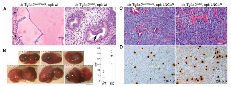
Transforming growth factor (TGF)-beta is an important paracrine factor in tumorigenesis. Ligand binding of the type I and II TGF-beta receptors initiate downstream signaling. The role of stromal TGF-beta signaling in prostate cancer progression is unknown. In mice, the conditional stromal knockout of the TGF-beta type II receptor expression (Tgfbr2(fspKO)) resulted in the development of prostatic intraepithelial neoplasia and progression to adenocarcinoma within 7 months. Clinically, we observed a loss of TGF-beta receptor type II expression in 69% of human prostate cancer-associated stroma, compared to 15% of stroma associated with benign tissues (n=140, P-value <0.0001). To investigate the mechanism of paracrine TGF-beta signaling in prostate cancer progression, we compared the effect of the prostatic stromal cells from Tgfbr2(fspKO) and floxed TGF-beta type II receptor Tgfbr2(floxE2/floxE2) mice on LNCaP human prostate cancer cells in vitro and tissue recombination xenografts. Induction of LNCaP cell proliferation and tumorigenesis was observed by Tgfbr2(fspKO) prostate stroma as a result of elevated Wnt3a expression. Neutralizing antibodies to Wnt3a reversed LNCaP tumorigenesis. The TGF-beta inhibition of Wnt3a expression was in part through the suppression of Stat3 activity on the Wnt3a promoter. In conclusion, the frequent loss of stromal TGF-beta type II receptor expression in human prostate cancer can relieve the paracrine suppression of Wnt3a expression.


Figures

Figure 1
Figure 1
The loss of TβRII expression in the prostatic stroma of mice leads to transformation of adjacent epithelia. A. Histologic comparison of Tgfbr2floxE2/floxE2 (left) and Tgfbr2fspKO (right) mouse prostates by H&E staining suggest Tgfbr2fspKO mouse prostates develop PIN by 6 weeks of age. B. Electron microscopy indicates the absence of secretory vesicles in Tgfbr2fspKO mouse prostates compared to their presence in Tgfbr2floxE2/floxE2 prostates. Scale bar represents 2 μm. C. Following tissue rescue of Tgfbr2floxE2/floxE2 and Tgfbr2fspKO prostates for seven months, the histology of the Tgfbr2fspKO prostates progressed to adenocarcinoma while the Tgfbr2floxE2/floxE2 prostates maintained a wild type phenotype. D. Immunohistochemistry for TβRII expression of the rescued tissues showed positive stromal and epithelial staining in the Tgfbr2floxE2/floxE2 prostates, yet only epithelial staining in the Tgfbr2fspKO prostates. Scale bar represents 50 μm for panels A, C, and D; 25 μm for insets.
Figure 2
Figure 2
Tgfbr2fspKO prostates develop adenocarcinoma in seven months. A. Phosphorylated-histone H3 staining of the mitotic cells suggested higher proliferation rate in the Tgfbr2fspKO prostates rescues than the normal Tgfbr2floxE2/floxE2 prostates rescues. The mean ± standard deviation of positive staining is indicated in each panel (P < 0.01, n = 6 for both test and control). B. Immunohistochemistry for mouse dorsolateral prostate secretory proteins (mDLP) was detectible control Tgfbr2floxE2/floxE2 prostates, but often lost in the Tgfbr2fspKO tissue rescues. C. Immunohistochemistry for p63 revealed disorganized staining pattern in the prostates of Tgfbr2fspKO compared to Tgfbr2floxE2/floxE2. D. Immunohistochemistry for Twist expression was positive in the adenocarcinoma Tgfbr2fspKO tissues, but not expressed in the Tgfbr2floxE2/floxE2 prostates. The sections were nuclear counterstained with hematoxylin (blue). Scale bar represents 50 μm for panels and 25 μm for insets.
Figure 3
Figure 3
Tgfbr2fspKO prostatic stromal cells have elevated Wnt3a expression. A. The screening of 19 Wnt isoforms by real-time PCR revealed specific Wnt isoforms to have greater mRNA expression by cultured Tgfbr2fspKO prostatic stromal cells relative to control, Tgfbr2floxE2/floxE2 stromal cells. Each dot represents a comparative expression level of a Tgfbr2fspKO sample relative to the average expression level of the Tgfbr2floxE2/floxE2 samples (baseline). The data were normalized to 18s ribosomal RNA expression. The dotted horizontal line is at the value of 1 representing no difference from the Tgfbr2floxE2/floxE2 average. The thick horizontal lines indicate the medians within each group. (There are 3 data (83.9, 88.0, 410.1) in Wnt3a and 1 (51.5) in Wnt10b that are out of the plot range.) B. Western blot confirmed specifically Wnt3a protein expression was greater in Tgfbr2fspKO prostatic stromal cells compared to that from Tgfbr2floxE2/floxE2 cells.
Figure 4
Figure 4
Wnt3a expression is down regulated by TGF-β in prostate stromal cells. A. Conditioned media from Tgfbr2floxE2/floxE2 or Tgfbr2fspKO stromal cells were incubated with LNCaP cells in the presence or absence of Wnt3a neutralizing antibody and isotype control IgG. Subsequently, luciferase reporter assay for the c-myc promoter was used as readout for canonical Wnt activity in LNCaP cells. B. Cell counting was used to measure LNCaP cell proliferation following incubation with Tgfbr2floxE2/floxE2 or Tgfbr2fspKO prostatic stromal conditioned media. The addition of Wnt3a neutralizing antibody inhibited LNCaP cell proliferation in a dose dependent manner. The graphs in panels A and B indicate mean ± standard deviation (P < 0.01, n = 12). C. Wnt3a promoter analysis suggested Stat transcription factor binding consensus sites. ChIP analysis suggested greater Stat3 binding to the Wnt3a -854 and -3542 promoter elements in Tgfbr2fspKO stromal cells compared to Tgfbr2floxE2/floxE2 stromal cells. D. Western blotting for Wnt3a following siRNA-knockdown of Stat3 in Tgfbr2fspKO stromal cells showed inhibition of Wnt3a expression. The scrambled (Scmb.) siRNA did not affect Wnt3a expression. Actin expression served as a loading control.
Figure 5
Figure 5
Tgfbr2fspKO prostatic stromal cells increase tumorigenicity of prostate epithelial cells. A. Tissue recombinant of Tgfbr2floxE2/floxE2 or Tgfbr2fspKO mouse prostate stromal cells with wild type mouse prostate organoids recapitulated the histology of the respective intact mice. Arrow indicates PIN lesion development in the tissue recombinant associated with Tgfbr2fspKO stromal cells. Asterisks indicate kidney parenchyma. B. The gross representations of the LNCaP/Tgfbr2fspKO tumors in renal xenografts were larger than control, LNCaP/Tgfbr2floxE2/floxE2 tumors. Tumor volumes calculated using Image J software were graphed as mean ± standard deviation (P < 0.01, n = 6). Scare bar represents 4 mm. C. H&E for the LNCaP/Tgfbr2floxE2/floxE2 and LNCaP/Tgfbr2fspKO recombinant tumors histology showed little difference. D. Immunohistochemistry for phosphorylated-histone H3, indicated the mitotic index of LNCaP/Tgfbr2fspKO tumors to be greater than LNCaP/Tgfbr2floxE2/floxE2 tumors. The mean positive staining is indicated in each panel ± standard deviation (P < 0.01, n = 6). The scale bar in panel A represents 50 μm for panels A, C, and D.
Figure 6
Figure 6
Wnt3a neutralizing antibody inhibits tumorigenic progression of LNCaP/Tgfbr2fspKO tissue recombinants. Wnt3a neutralizing antibody or isotype control IgG was i. p. injected to the hosted SCID mice twice a week for two weeks, started two weeks post-grafting. A. Histology by H&E showed increased necrotic areas in the tumors from Wnt3a antibody injected mice, compared to the ones from control mice (indicated by dashed line). B. Greater number of apoptotic cells was localized in the tumors treated with Wnt3a neutralizing antibody compared to control by Apop-tag immunohistochemistry (P < 0.01, n = 8). C. Immunohistochemistry of phosphorylated-histone H3 showed less mitotic cells in the tumors from Wnt3a treated mice compared to control (P < 0.01, n = 6). The mean ± standard deviation is indicated in each panel. The scale bar represents 50 μm for panels and 25 μm for insets.
Figure 7
Figure 7
Immunohistochemistry for TGF-β type II receptor (TβRII) expression is not detectable in stromal cells of human prostate adenocarcinomas. The pathologic grade of the representative immunohistochemistry images is indicated as benign or Gleason score. Note TβRII was consistently expressed in epithelial cells, but often lost in stromal cells of neoplastic tissues. Scale bar represents 50 μm. The table indicates the distribution of tissue pathology with positive histochemical TβRII staining in the stromal compartment.

References

    1. Bhowmick NA, Chytil A, Plieth D, Gorska AE, Dumont N, Shappell S, et al. TGF-beta signaling in fibroblasts modulates the oncogenic potential of adjacent epithelia. Science. 2004a;303:848–51. - PubMed
    1. Bhowmick NA, Moses HL. Tumor-stroma interactions. Curr Opin Genet Dev. 2005;15:97–101. - PMC - PubMed
    1. Bhowmick NA, Neilson EG, Moses HL. Stromal fibroblasts in cancer initiation and progression. Nature. 2004b;432:332–7. - PMC - PubMed
    1. Bierie B, Moses HL. TGF-beta and cancer. Cytokine Growth Factor Rev. 2006;17:29–40. - PubMed
    1. Biswas S, Guix M, Rinehart C, Dugger TC, Chytil A, Moses HL, et al. Inhibition of TGF-beta with neutralizing antibodies prevents radiation-induced acceleration of metastatic cancer progression. J Clin Invest. 2007;117:1305–13. - PMC - PubMed

Publication types

MeSH terms