. 2008 Aug 22;134(4):599-609.
doi: 10.1016/j.cell.2008.06.033.

A sequence motif within chromatin entry sites directs MSL establishment on the Drosophila X chromosome

Affiliations

A sequence motif within chromatin entry sites directs MSL establishment on the Drosophila X chromosome

Artyom A Alekseyenko et al. Cell. .

Abstract

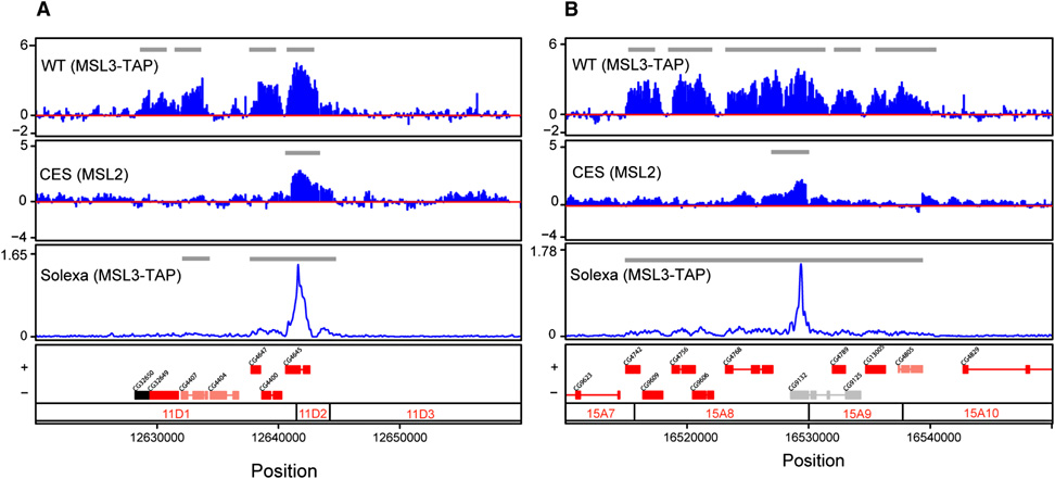
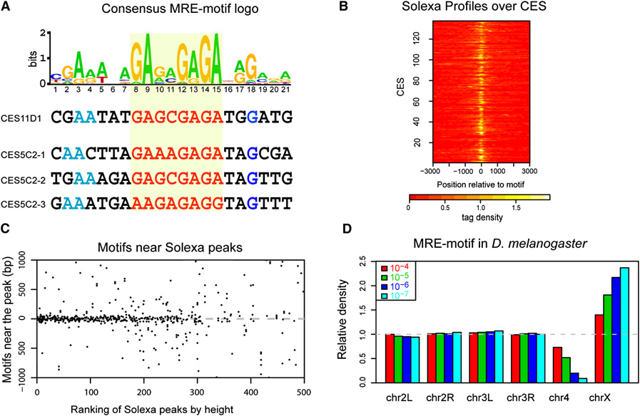
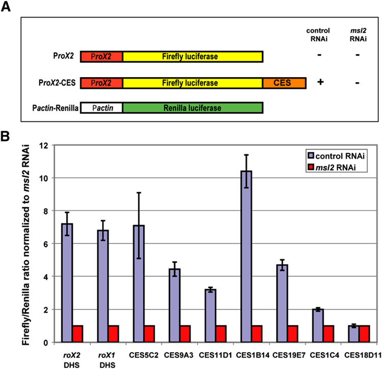
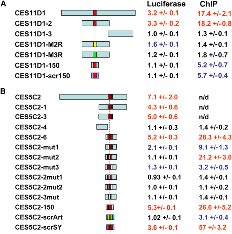
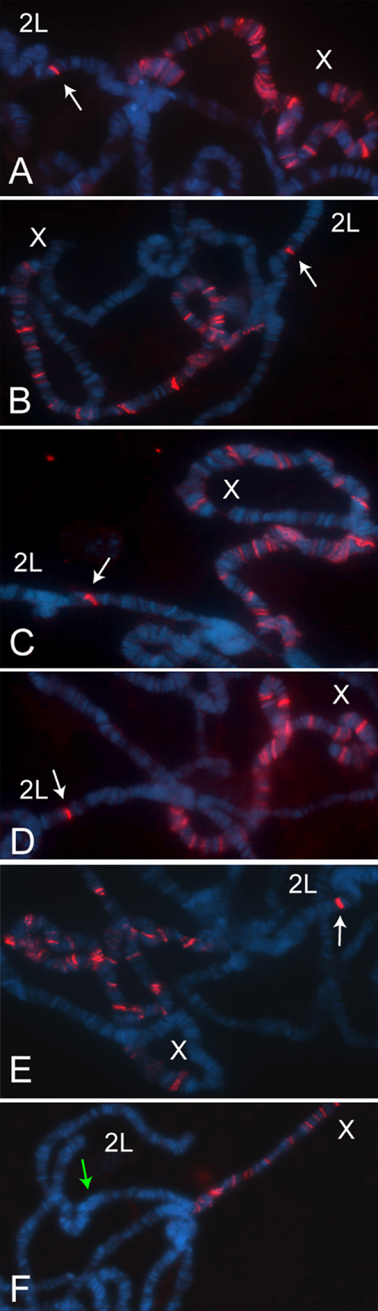
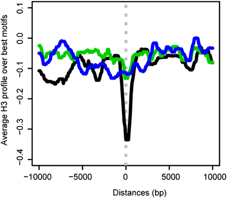
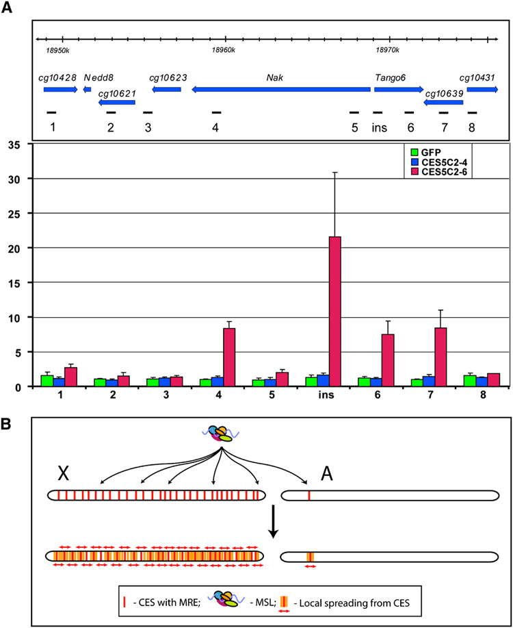
The Drosophila MSL complex associates with active genes specifically on the male X chromosome to acetylate histone H4 at lysine 16 and increase expression approximately 2-fold. To date, no DNA sequence has been discovered to explain the specificity of MSL binding. We hypothesized that sequence-specific targeting occurs at "chromatin entry sites," but the majority of sites are sequence independent. Here we characterize 150 potential entry sites by ChIP-chip and ChIP-seq and discover a GA-rich MSL recognition element (MRE). The motif is only slightly enriched on the X chromosome ( approximately 2-fold), but this is doubled when considering its preferential location within or 3' to active genes (>4-fold enrichment). When inserted on an autosome, a newly identified site can direct local MSL spreading to flanking active genes. These results provide strong evidence for both sequence-dependent and -independent steps in MSL targeting of dosage compensation to the male X chromosome.


Figures

Figure 1
Figure 1. MSL3-independent chromatin entry sites are a subset of the wild-type binding pattern for MSL complex and coincide with the strongest enrichment peaks detected by ChIP-seq
Two representative chromatin entry sites, CES11D1 (A) and CES15A8 (B) are shown. ChIP-chip profiles were generated from y w; MSL3-TAP; msl3 embryonic chromatin (WT) using IgG to IP the TAP epitope, or msl3 mutant embryos (CES) using anti-MSL2 antibodies. DNA resulting from ChIP was hybridized to custom NimbleGen tiling arrays (Alekseyenko et al., 2006). The y axis shows the log2 ratio of IP/Input signal. The ChIP-seq tag profile (Solexa) was obtained from an MSL3-TAP transformed male cell line, Clone8, using IgG to IP the TAP epitope. The ChIP-seq profile displays broad distribution along WT MSL targets and high peaks that correspond to entry sites. The y axis shows the tag density. Gray lines within ChIP-chip and ChIP-seq panels indicate the regions identified as bound clusters (See Experimental Procedures for details). Genes are color coded based on their transcriptional status (transcribed, red; nontranscribed, black; genes that are differentially transcribed in S2 and Clone 8 cells, salmon; and genes without transcriptional data, gray). Genes on the top row are transcribed left to right, and genes on the bottom row are transcribed from right to left. Numbers along the x axis refer to chromosomal position (bp) (Dm1 release coordinates). Polytene map cytological locations are indicated below.
Figure 2
Figure 2. A GA-rich motif is a common feature in MSL chromatin entry sites
(A) Identification of the MRE-motif enriched in CES. The motif logo is shown with representative examples below. This motif occurs once in CES11D1 and three times in CES5C2. The GA-rich core is highlighted in red. We are arbitrarily showing the GA-rather than the TC-rich strand. This motif was identified using the chromatin entry sites as determined by the ChIP-chip data; a nearly identical motif is obtained from the WT Solexa data (Suppl. Fig. 2S). (B) Solexa peaks center on the MRE-motif in chromatin entry sites. Shown is a heatmap of Solexa profiles (tag densities increase from red to white), aligned by the MRE-motif in each of the 137 individual CES that contains the motif. In nearly all cases, the location of the sharp peaks coincides with the motif location. (C) The MRE-motif tends to localize to the center of strong Solexa peaks. The top 500 Solexa peaks were ranked by height on the x axis and then MRE-motifs within a 2 kb window flanking each peak were mapped on the y axis. Some peaks contain multiple motifs; to avoid showing the same region twice, only the strongest peaks in any 3 kb region were considered. Most motifs map near the center, but motif centering decreases as peak intensity weakens. (D) The MRE-motif is enriched on the X chromosome, compared to autosomes. Motif frequency for each chromosome was normalized to its frequency on all autosomes. Colors correspond to different thresholds for defining the occurrence of the motif. At all thresholds, the motif is enriched on the X. As the stringency increases, the enrichment increases but a smaller number of motifs are identified.
Figure 3
Figure 3. Test for CES function in a luciferase reporter assay
(A) Experimental design, and structure of the constructs used for a dual Firefly/Renilla luciferase reporter assay. S2 cells co-transfected with the firefly luciferase construct containing each 1.5 kb CES fragment and the control Renilla luciferase plasmid were treated with dsRNA against either GFP (control) or MSL2 (experimental). (B) Examples of Firefly/Renilla luciferase activity conferred by nine chromatin entry sites normalized to the value obtained after msl2 RNAi. CES18D11 is a rare example of a CES that failed to show MSL2-dependent upregulation in this assay.
Figure 4
Figure 4. The MRE-motif is required for MSL complex binding to CES
Structure of the DNA fragments tested in the context of CES11D1 (A) and CES5C2 (B), and the activities they display in the luciferase assay in S2 cells and when inserted in a specific location (37B7) on chromosome 2L in transgenic flies (MSL2 ChIP). Numerical values in red were scored positive, black were negative, and blue were considered intermediate. Positions of the motifs are shown as red boxes; when mutated, MRE-motifs are shown as non-red boxes (please refer to Suppl. Fig. 3S for sequence changes within the motif). Randomized DNA sequences in the shortest subclones not affecting the MRE-motifs are shown in purple or dark blue (with only the motif core remaining intact). Luciferase and ChIP data are based on at least three independent experiments. n/d – not determined.
Figure 5
Figure 5. MSL complex is recruited to entry sites placed on autosomes
Polytene chromosomes from homozygous CES-X{37B7}; H83M2-6I, msl3 females were immunostained with anti-MSL1 antibodies. (A) roX1 DHS (300bp). (B) CES11D1 (1.5 kb). (C) CES9A3 (1.5 kb). (D) CES5C2 (1.5 kb). (E) CES5C2-6 (268 bp, containing three motifs). (F) CES5C2-4 (268 bp, lacking a motif). White arrows point to the positive signal at the integration site, a green arrow points to the cytological location with no visible staining. The X chromosome in each nucleus displays the partial pattern typical of msl3 mutants.
Figure 6
Figure 6. Histone H3 is depleted over MREs in chromatin entry sites
Average H3 profile centered over the best MRE within 137 CES (black) shows clear depletion. In contrast, the best (green) or a random set (blue) of 137 MRE-motifs outside CES display a relatively flat profile. The histone occupancy data are from S2 and Clone8 cells (Larschan et al., 2007).
Figure 7
Figure 7. MSL complex spreads into neighboring genes from CES5C2 inserted on an autosome
(A) MSL3-TAP ChIP in the region surrounding the 37B7 integration site where either GFP (1.9 kb), CES5C2-4 (258 bp, no MREs) or CES5C2-6 (268 bp, three MREs) are placed (y w; CES-X{37B7}/ MSL3-TAP; H83M2-6I, msl3 mixed sex embryos). Top panel: genetic map and location of probes, Dm3 release coordinates are shown. Bottom panel: MSL3-TAP data for GFP (green), CES5C2-4 (blue) and CES5C2-6 (red), indicate that only CES5C2-6 effectively targets the MSL complex and directs limited spreading to neighboring genes. (B) Two-step model for MSL targeting of the X chromosome. X, X chromosome, A, autosome. First step: MSL complex targets >150 chromatin entry sites containing MRE motifs on the X chromosome. The autosome is normally ignored, unless a CES from the X is inserted on the autosome. Second step: Local spreading from entry sites leads to MSL binding to the majority of active genes on the X chromosome. In addition, an ectopic CES can lead to targeting of flanking active genes on an autosome, as seen in the results displayed in panel A.

References

    1. Alekseyenko AA, Larschan E, Lai WR, Park PJ, Kuroda MI. High-resolution ChIP-chip analysis reveals that the Drosophila MSL complex selectively identifies active genes on the male X chromosome. Genes Dev. 2006;20:848–857. - PMC - PubMed
    1. Amrein H, Axel R. Genes expressed in neurons of adult male Drosophila. Cell. 1997;88:459–469. - PubMed
    1. Bai X, Alekseyenko AA, Kuroda MI. Sequence-specific targeting of MSL complex regulates transcription of the roX RNA genes. EMBO Journal. 2004;23:2853–2861. - PMC - PubMed
    1. Bailey TL, Williams N, Misleh C, Li WW. MEME: discovering and analyzing DNA and protein sequence motifs. Nucleic Acids Res. 2006;34:W369–W373. - PMC - PubMed
    1. Bashaw GJ, Baker BS. The msl-2 dosage compensation gene of Drosophila encodes a putative DNA-binding protein whose expression is sex specifically regulated by Sex-lethal. Development. 1995;121:3245–3258. - PubMed

Publication types

Associated data