. 2008 Aug 22;134(4):634-45.
doi: 10.1016/j.cell.2008.06.025.

The GET complex mediates insertion of tail-anchored proteins into the ER membrane

Affiliations

The GET complex mediates insertion of tail-anchored proteins into the ER membrane

Maya Schuldiner et al. Cell. .

Abstract

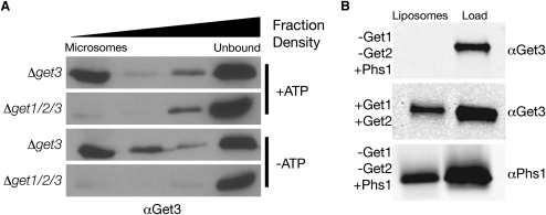
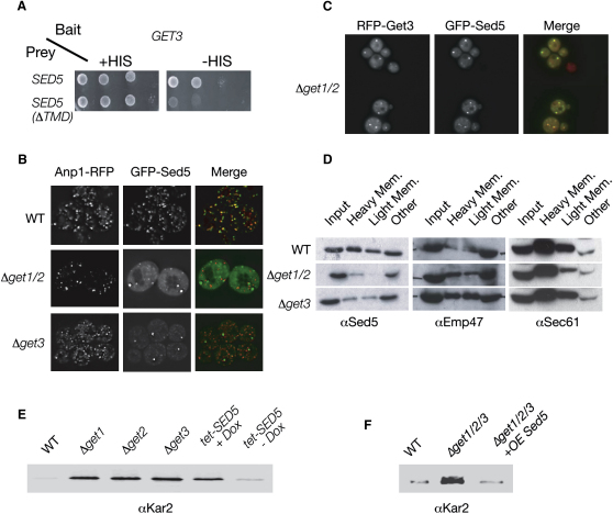
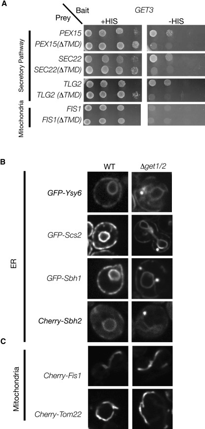
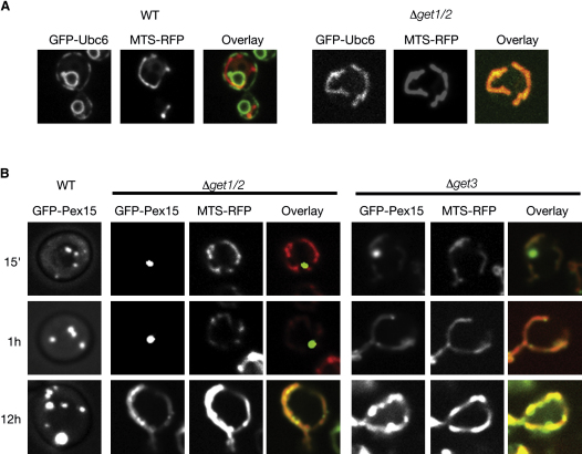
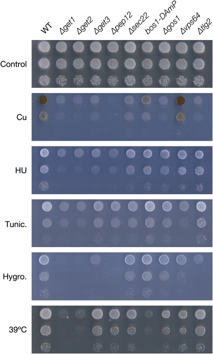
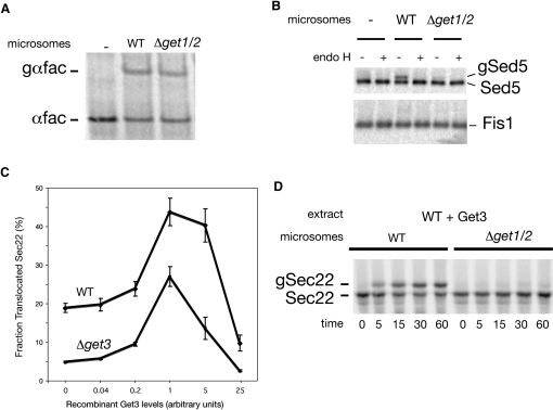
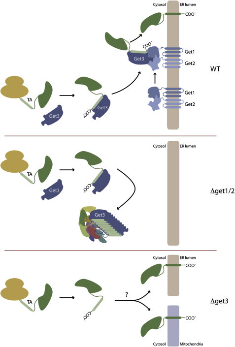
Tail-anchored (TA) proteins, defined by the presence of a single C-terminal transmembrane domain (TMD), play critical roles throughout the secretory pathway and in mitochondria, yet the machinery responsible for their proper membrane insertion remains poorly characterized. Here we show that Get3, the yeast homolog of the TA-interacting factor Asna1/Trc40, specifically recognizes TMDs of TA proteins destined for the secretory pathway. Get3 recognition represents a key decision step, whose loss can lead to misinsertion of TA proteins into mitochondria. Get3-TA protein complexes are recruited for endoplasmic reticulum (ER) membrane insertion by the Get1/Get2 receptor. In vivo, the absence of Get1/Get2 leads to cytosolic aggregation of Get3-TA complexes and broad defects in TA protein biogenesis. In vitro reconstitution demonstrates that the Get proteins directly mediate insertion of newly synthesized TA proteins into ER membranes. Thus, the GET complex represents a critical mechanism for ensuring efficient and accurate targeting of TA proteins.


Figures

Figure 1
Figure 1
Get1 and Get2 Act as a Membrane Receptor for the Soluble Get3 (A) Western blots with αGet3 showing binding of recombinant Get3 ATPase to microsomes prepared from Δget3 or Δget1/2/3 strains in the presence or absence of ATP. Shown are Optiprep gradient fractions, which separate microsomes from unbound protein. (B) Western blots with αGet3 or αPhs1 showing binding of recombinant Get3 to proteoliposomes reconstituted with either Phs1 as a control protein (−Get1/−Get2+PHS1) or purified Get1-PC and Get2-HA (+Get1/+Get2). Shown are optiprep gradient fractions as above.
Figure 2
Figure 2
Get3 Binds to Sed5 and Is Important for Its Biogenesis In Vivo (A) Yeast two-hybrid assay with Get3 as bait and Sed5197–340 (the strongest hit from the Y2H screen) as prey (in the presence or absence of its TMD). The growth on medium lacking histidine (−HIS) is indicative of a physical interaction. (B) Fluorescence microscopy demonstrating a shift in the subcellular localization of GFP-Sed5 from Golgi in control (WT) strains to a partially cytosolic localization in a Δget3 strain, and both cytosolic and few large puncta in Δget1/2 strains. The GFP-Sed5 puncta in get mutants do not colocalize with the Golgi marker Anp1-RFP. (C) Fluorescence microscopy demonstrating colocalization of GFP-Sed5 and Get3-tdRFP in cytosolic aggregates that form in a Δget1/2 background. (D) Western blots of cell fractionation experiments to determine levels of Sed5 in membrane fractions. Control (WT), Δget1/2 or Δget3 strains were divided into three fractions (Heavy Mem, Lighter Mem, and remainder of cellular proteins [Other]) and compared to input protein (Input) with Western blots immunostained against either Sed5 or the control Golgi transmembrane protein, Emp47, and ER transmembrane protein Sec61. (E) Western blots of secreted proteins with αKar2. Assay for Kar2 secretion was performed on a control strain (WT), mutants of the GET complex (Δget1, Δget2, Δget3), and on a yeast strain harboring a repressible allele of the essential TA protein Sed5 (tet-SED5), either in the presence (+Dox) or absence (−Dox) of the corepressor doxycycline. (F) Western blots of secreted proteins with αKar2. Assay for Kar2 secretion was performed on the triple mutant (Δget1/2/3) either alone or overexpressing SED5 from a high copy plasmid (+ OE SED5), and compared to a control strain (WT).
Figure 3
Figure 3
The GET Complex Affects the Biogenesis of a Wide Variety of TA Proteins (A) Y2H assay showing Get3 as bait and various TA proteins (in the presence or absence of their TMDs) as prey. The growth on medium lacking histidine (−HIS) is indicative of a physical interaction. (B) Fluorescence microscopy of control (WT) and Δget1/2 strains expressing a broad variety of TA proteins. GFP-Scs2, GFP-Sbh1, and GFP-Ysy6 under a galactose-inducible (GAL) promoter. Cherry-Sbh2 was expressed from a plasmid under the constitutive TEF2 promoter. (C) Fluorescence microscopy of control (WT) and Δget1/2 strains expressing two mitochondrial TA proteins, Cherry-Fis1 and Cherry-Tom22, expressed from a plasmid under the constitutive TEF2 promoter.
Figure 4
Figure 4
Role of GET Proteins in Creating Membrane Specificity (A) Fluorescence microscopy showing the localization of GFP-Ubc6 and mitochondrially targeted dsRED (MTS-RFP) in a control (WT) or Δget1/2 strain. (B) Fluorescence microscopy of a time course monitoring the subcellular localizations of the peroxisomal TA protein GFP-Pex15 as well as dsRED targeted to the mitochondria (MTS-RFP) following induction of Pex15 from a galactose inducible promoter in a control (WT), get1/2, or get3 strain.
Figure 5
Figure 5
Reduced Levels of TA Proteins Can Explain the Diverse Array of GET Complex Phenotypes Serial dilutions in different conditions: SD + CuSO4 (Cu), SD + hydroxyurea (HU), SD + tunicamycin (Tunic.), SD + hygromycin (Hygro.), and YPD incubated at 39°C (39°C). Strains shown are: control cells (WT), get mutants, five TA protein deletion strains, and a strain carrying a hypomorphic allele (DAmP) of an essential TA protein. Copper sensitivity in the Δget3 strain is more pronounced in methionine prototrophic than auxotrophic cells. We used Δmet15 cells for this panel, resulting in a less sensitive phenotype compared with MET+ cells depicted in Figure S4.
Figure 6
Figure 6
In Vitro Reconstitution of GET-Dependent Insertion of TA Proteins (A) Autoradiograph of in vitro-translated, 35S methionine-labeled, α-factor (αfac) following incubation in the presence of microsomes derived from WT or Δget1/2 strains. The position of untranslocated prepro-αfac and glycosylated, translocated pro-αfac (gαfac) are indicated. (B) Autoradiograph of in vitro translated, 35S methionine-labeled Sed5 and Fis1 following incubation with microsomes derived from WT or Δget1/2 strains. Prior to SDS-PAGE analysis, samples were immunoprecipitated with an anti-opsin antibody and then treated with EndoH, as indicated. The position of untranslocated Sed5 and Fis1 as well as glycosylated translocated Sed5 (gSed5) are indicated. (C) Graph representing the dose dependence of Sec22 translocation on addition of recombinant Get3 to either WT- or Δget3-derived translation extracts. WT microsomes were added following translation, and the amount of glycosylated Sec22 relative to total Sec22 was calculated. Results from three independent experiments are shown; data are presented as mean ± SD. (D) Autoradiograph of in vitro-translated, 35S methionine labeled, Sec22 following translation in WT cytosol supplemented with optimal levels of Get3. Translocation was terminated at the indicated times following addition of microsomes derived from either WT or Δget1/2 strains. The position of untranslocated Sec22 as well as glycosylated translocated Sec22 (gSec22) are indicated.
Figure 7
Figure 7
Schematic Model for GET Complex Function (Top) WT cells. Get3 recognizes newly synthesized, ER-destined TA proteins. The Get3-TA complexes dock onto the Get1/Get2 receptor. This allows insertion of TA proteins. (Middle) Cells lacking the receptor (Δget1/2). Get3-TA complexes fail to reach the ER and, instead, are sequestered in cytosolic aggregates. (Bottom) Cells lacking Get3 (Δget3). Newly synthesized TA proteins intended for the ER are no longer shuttled into the GET pathway. To varying degrees, depending on the TA proteins, they may use alternate ATP/GTP-dependant pathways or spontaneous routes for membrane insertion. This could lead to misinsertion into the mitochondria, inefficient insertion into the ER, or aggregation in the cytosol.

References

    1. Abell B.M., Pool M.R., Schlenker O., Sinning I., High S. Signal recognition particle mediates post-translational targeting in eukaryotes. EMBO J. 2004;23:2755–2764. - PMC - PubMed
    1. Abell B.M., Rabu C., Leznicki P., Young J.C., High S. Post-translational integration of tail-anchored proteins is facilitated by defined molecular chaperones. J. Cell Sci. 2007;120:1743–1751. - PubMed
    1. Auld K.L., Hitchcock A.L., Doherty H.K., Frietze S., Huang L.S., Silver P.A. The conserved ATPase Get3/Arr4 modulates the activity of membrane-associated proteins in Saccharomyces cerevisiae. Genetics. 2006;174:215–227. - PMC - PubMed
    1. Banfield D.K., Lewis M.J., Rabouille C., Warren G., Pelham H.R. Localization of Sed5, a putative vesicle targeting molecule, to the cis-Golgi network involves both its transmembrane and cytoplasmic domains. J. Cell Biol. 1994;127:357–371. - PMC - PubMed
    1. Beilharz T., Egan B., Silver P.A., Hofmann K., Lithgow T. Bipartite signals mediate subcellular targeting of tail-anchored membrane proteins in Saccharomyces cerevisiae. J. Biol. Chem. 2003;278:8219–8223. - PubMed

Publication types

MeSH terms

Substances