. 2008 Nov;28(21):6609-19.
doi: 10.1128/MCB.00398-08. Epub 2008 Aug 25.

Embryonic stem cell-specific miR302-367 cluster: human gene structure and functional characterization of its core promoter

Affiliations

Embryonic stem cell-specific miR302-367 cluster: human gene structure and functional characterization of its core promoter

Alicia Barroso-delJesus et al. Mol Cell Biol. 2008 Nov.

Abstract

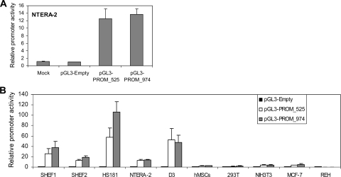
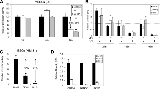
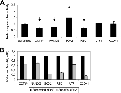
MicroRNAs (miRNAs) play a central role in the regulation of multiple biological processes including the maintenance of stem cell self-renewal and pluripotency. Recently, the miRNA cluster miR302-367 was shown to be differentially expressed in embryonic stem cells (ESCs). Unfortunately, very little is known about the genomic structure of miRNA-encoding genes and their transcriptional units. Here, we have characterized the structure of the gene coding for the human miR302-367 cluster. We identify the transcriptional start and functional core promoter region which specifically drives the expression of this miRNA cluster. The promoter activity depends on the ontogeny and hierarchical cellular stage. It is functional during embryonic development, but it is turned off later in development. From a hierarchical standpoint, its activity decays upon differentiation of ESCs, suggesting that its activity is restricted to the ESC compartment and that the ESC-specific expression of the miR302-367 cluster is fully conferred by its core promoter transcriptional activity. Furthermore, algorithmic prediction of transcription factor binding sites and knockdown studies suggest that ESC-associated transcription factors, including Nanog, Oct3/4, Sox2, and Rex1 may be upstream regulators of miR302-367 promoter. This study represents the first identification, characterization, and functional validation of a human miRNA promoter in stem cells. This study opens up new avenues to further investigate the upstream transcriptional regulation of the miR302-367 cluster and to dissect how these miRNAs integrate in the complex molecular network conferring stem cell properties to ESCs.


Figures

FIG. 1.
FIG. 1.
Heat map representation of relative miRNA expression profiling. Expression levels of a panel of 170 miRNAs were determined by quantitative real-time RT-PCR. Data were normalized against GAPDH mRNA levels, and the expression level is relative to the adult tissue mix reference sample. Pure green spots represent at least 20-fold downregulation of the miRNA in the cell line under study in comparison to the reference sample. Pure red spots indicate at least 20-fold upregulation of the miRNA in the sample under study in comparison to the reference. Black spots indicate no difference in the miRNA expression level in the sample compared to the reference. Heat map representation and data clustering were performed with Cluster and Treeview software.
FIG. 2.
FIG. 2.
Intracellular distribution of miR302 family members. Northern blot analysis was used to determine the nuclear versus cytoplasmic abundance of miR302a, miR302b, miR303c, and miR302d. All membranes were reprobed with U6 snRNA and tRNA-Lys to verify proper RNA fractionation.
FIG. 3.
FIG. 3.
Transcriptional unit of miR302-367 gene. (A) 5′ RACE assay shows specific PCR amplification only in the presence of RT and TAP treatment (lane 2). Identical reactions without TAP treatment (lane 3) or without RT (lane 4) gave no amplification. M, molecular marker. (B) Sequence diagram depicting the gene structure and main genomic components of the human miR302-367 transcriptional unit. The first nucleotide to be transcribed is numbered as +1 (transcription start). Canonical sequence motifs for TATA box and polyadenylation signal are boxed. Intron sequences are shown in brackets and shortened due to their extension. The miR302-367 cluster is located within the first intron. (C) Adapted 3′ RACE assay displaying specific PCR product. No band was observed in the absence of RT. Two bands illustrated by arrows were clearly detectable. (D) Detection of putative polyadenylated unspliced pri-miRNA by Northern blot analysis in NTERA-2. Total RNA (7 μg) was loaded in lane 1. From the same amount of total RNA, poly(A) (lane 2) and poly(A)+ (lane 3) fractions were loaded.
FIG. 4.
FIG. 4.
Representation displaying the structure homology of the miR302-367 locus. The upper part of the figure depicts a schematic representation of the human miR302-367 locus. The transcriptional start (+1) is shown. Black arrows represent the miRNA precursor. Gray boxes represent the miR302-369 exons. Lined boxes indicate the introns. The polyadenylation signal is represented as gray vertical lines. The conserved promoter sequence is identified upstream of the transcriptional start as a dotted arrow. White arrows represent LARP7 exons. Alternative splicing of the primary RNA is also shown. The lower part of the figure shows an overall profile of sequence similarity (represented in red) obtained from the multiple alignment of H. sapiens (hs), M. musculus (mm), C. familiaris (cf), and B. taurus (bt) homologues. Multiple sequence alignment was performed using the Lagan program of Vista tools.
FIG. 5.
FIG. 5.
Transcriptional activity of genomic regions PROM_525 and PROM_974. (A) Both genomic regions were PCR amplified from the hECC line NTERA-2 and subcloned into the promoterless luciferase reporter vector pGL3Basic (Promega). Either the pGL3-PROM_525 or pGL3-PROM_974 construct was cotransfected along with a Renilla reporter plasmid into the hECC line NTERA-2. Luciferase activity was measured 24 h after transfection. Luciferase activity was normalized against Renilla activity and expressed as promoter activity relative to the empty vector (pGL3-Empty). Data from three independent experiments are represented as means ± standard deviations. (B) The transcriptional activity of both the 525-bp and the 974-bp motifs was determined in multiple cell types at different ontogenetic stages of development: genetically stable hESC lines SHEF1, SHEF2, and HS181; the transformed hECC line NTERA-2; and mESC line D3 are ESC lines. hMSCs are adult MSCs. In addition to these primary cells with stem cell potential, four transformed cell lines with no stem cell capacity were studied: the human embryonic kidney cell line 293T, the murine embryonic fibroblastic cell line NIH 3T3, and the human adult breast line MCF-7 and B-cell leukemic line REH. Data are presented as described for panel A.
FIG. 6.
FIG. 6.
Loss of promoter transcriptional activity upon ESC differentiation. (A) The construct pGL3-PROM_974 and the Renilla reporter were cotransfected in mESCs. Undifferentiated mESCs (UnESC) were maintained on gelatin-coated plates in the presence of LIF (solid black bars). mESCs were differentiated through embryoid body formation in the absence (open bars) or presence (gray bars) of RA. The promoter activity was assessed at 24, 48, and 96 h after transfection. Luciferase activity was normalized against Renilla. The promoter activity is expressed relative to the undifferentiated mESCs. (B) Real-time RT-PCR analysis for the ESC-specific transcription factors Oct3/4, Nanog, and Rex1. Data acquisition and analysis were performed using the Mx3500P device (Stratagene). RNA expression is normalized against β-actin and relative to undifferentiated ESCs, which were used as the calibrator. dR, baseline-corrected raw fluorescence data. The dotted line defines the relative RNA expression of the calibrator sample. (C) hESCs were differentiated by prolonged culture on Matrigel-coated plates in the presence of differentiation medium (80% KO DMEM, 20% FBS, 2 mM l-glutamine, 2 mM nonessential amino acids, 50 μg/ml penicillin-streptomycin, and 10 nM β-mercaptoethanol, supplemented with 10 μM RA and 50 ng/ml of BMP4). Cells were assayed for PROM_974 activity at days 0, 4, and 7 after induction of differentiation (Undif, Dif 4d, and Dif 7d, respectively). Cells were cotransfected with construct pGL3-PROM_974 and the Renilla reporter 24 h before promoter activity measurements. Promoter activity is expressed relative to the undifferentiated hESCs (day 0). ↓, potential transcriptional activators; *, potential transcriptional repressor. (D) Real-time RT-PCR analysis for the ESC-specific transcription factors. Data acquisition and analysis were performed using the Mx3500P device (Stratagene). RNA expression is normalized against GAPDH and relative to undifferentiated hESCs (Undif), which were used as the calibrator.
FIG. 7.
FIG. 7.
Regulation of the miR302-367 promoter activity by ESC transcription factors. (A) The activity of the miR302-367 promoter (PROM_974 region) was evaluated after siRNA-mediated knockdown of potential regulating transcription factors: Oct3/4, Nanog, Rex1, and Sox2. Two additional ESC-associated transcription factors were tested: UTF-1 and CCDN1. Cells were cotransfected with the pGL3-PROM_974 construct, the Renilla reporter plasmid, and the corresponding siRNA. A scrambled siRNA with no homology to mouse or human sequences was used as a control. Measurements were performed 24 h posttransfection. Normalized promoter activity is expressed relative to scrambled siRNA-transfected cells (dotted line). The mean ± standard deviation from three independent experiments is shown. (B) siRNA-mediated inhibition of the transcription factors studied was confirmed by real-time PCR 24 h posttransfection. Data were normalized to GAPDH. Scrambled siRNA-transfected cells were used as a calibrator sample. dR, baseline-corrected raw fluorescence data.

References

    1. Ambros, V. 2004. The functions of animal miRNAs. Nature 435350-355. - PubMed
    1. Aukerman, M. J., and H. Sakai. 2003. Regulation of flowering time and floral organ identity by a microRNA and its APETALA2-like target genes. Plant Cell 152730-2741. - PMC - PubMed
    1. Bartel, D. P. 2004. MicroRNAs: genomics, biogenesis, mechanism, and function. Cell 116281-297. - PubMed
    1. Bernstein, E., S. Y. Kim, M. A. Carmell, E. P. Murchison, H. Alcorn, M. Z. Li, A. A. Mills, S. J. Elledge, K. V. Anderson, and G. J. Hannon. 2003. Dicer is essential for mouse development. Nat. Genet. 35215-217. - PubMed
    1. Borchert, G., W. Lanier, and B. Davidson. 2006. RNA polymerase III transcribes human microRNAs. Nat. Struct. Mol. Biol. 131097-1101. - PubMed

Publication types

MeSH terms