. 2008 Sep 1;205(9):1967-73.
doi: 10.1084/jem.20071632. Epub 2008 Aug 25.

Stimulation of Toll-like receptor 3 and 4 induces interleukin-1beta maturation by caspase-8

Affiliations

Stimulation of Toll-like receptor 3 and 4 induces interleukin-1beta maturation by caspase-8

Jonathan Maelfait et al. J Exp Med. .

Abstract

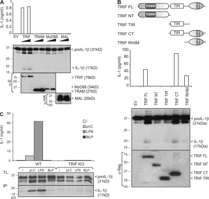
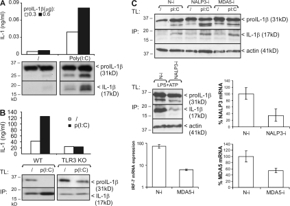
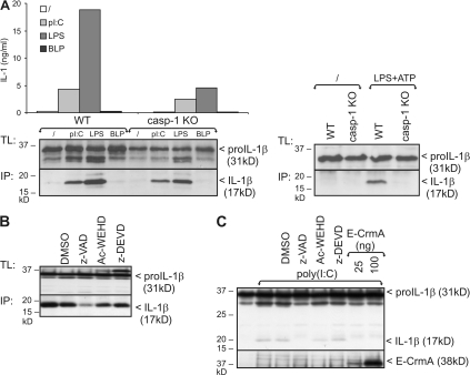
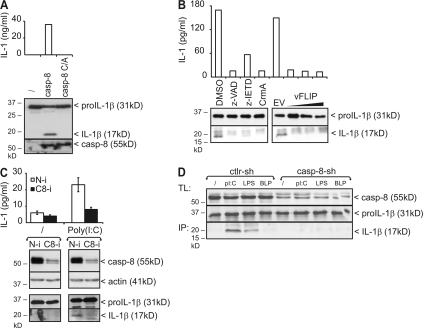
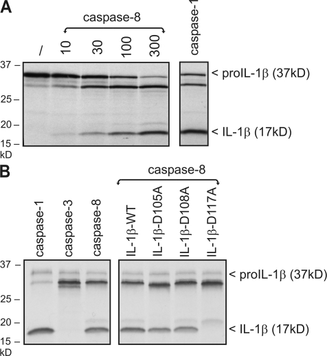
The cytokine interleukin (IL)-1beta is a key mediator of the inflammatory response and has been implicated in the pathophysiology of acute and chronic inflammation. IL-1beta is synthesized in response to many stimuli as an inactive pro-IL-1beta precursor protein that is further processed by caspase-1 into mature IL-1beta, which is the secreted biologically active form of the cytokine. Although stimulation of membrane-bound Toll-like receptors (TLRs) up-regulates pro-IL-1beta expression, activation of caspase-1 is believed to be mainly initiated by cytosolic Nod-like receptors. In this study, we show that polyinosinic:polycytidylic acid (poly[I:C]) and lipopolysaccharide stimulation of macrophages induces pro-IL-1beta processing via a Toll/IL-1R domain-containing adaptor-inducing interferon-beta-dependent signaling pathway that is initiated by TLR3 and TLR4, respectively. Ribonucleic acid interference (RNAi)-mediated knockdown of the intracellular receptors NALP3 or MDA5 did not affect poly(I:C)-induced pro-IL-1beta processing. Surprisingly, poly(I:C)- and LPS-induced pro-IL-1beta processing still occurred in caspase-1-deficient cells. In contrast, pro-IL-1beta processing was inhibited by caspase-8 peptide inhibitors, CrmA or vFLIP expression, and caspase-8 knockdown via RNAi, indicating an essential role for caspase-8. Moreover, recombinant caspase-8 was able to cleave pro-IL-1beta in vitro at exactly the same site as caspase-1. These results implicate a novel role for caspase-8 in the production of biologically active IL-1beta in response to TLR3 and TLR4 stimulation.


Figures

Figure 1.
Figure 1.
Poly(I:C) and LPS induce pro–IL-1β processing via a TRIF-dependent signaling pathway. (A) HEK293T cells were cotransfected with pro–IL-1β and 50 or 100 ng of either E-TRIF, E-TRAM, E-MyD88, or HA-MAL. 24 h later, pro–IL-1β processing and expression of transfected proteins was analyzed by Western blotting of total cell lysates (bottom). Secretion of biologically active IL-1β into the corresponding cell supernatants was analyzed via IL-1 bioassay (top). (B) HEK293T cells were cotransfected with pro–IL-1β and different Flag-tagged TRIF deletion mutants. 24 h later, pro–IL-1β processing and secretion of biologically active IL-1β was analyzed as in A. Expression of TRIF mutants was verified by Western blotting and detection with anti-Flag. (top) Schematic representation of the different TRIF deletion mutants. FL, full length; NT, N-terminal fragment; CT, C-terminal fragment; PEEMSW, TRAF6-binding motif; TIR, Toll/IL-1 receptor domain. (C) BLP-primed peritoneal macrophages from WT and TRIF KO mice were incubated for 18 h with poly(I:C), LPS, BLP, or control medium, as described in the Materials and methods. Pro–IL-1β was analyzed in total cell lysates (TL) by Western blotting (bottom). Mature IL-1β was detected in the cell supernatant upon IL-1β immunoprecipitation (IP). Secretion of biologically active IL-1β was analyzed via IL-1 bioassay (top). Data are representative of three independent experiments.
Figure 2.
Figure 2.
Poly(I:C)-induced pro–IL-1β processing is TLR3 dependent. (A) HEK293-TLR3 cells transfected with 0.3 or 0.6 μg pCAGGS-pro–IL-1β were incubated in the absence or presence of 25 μg/ml poly(I:C) for 6 h. Pro–IL-1β processing was analyzed by SDS-PAGE and Western blotting of total cell lysates (bottom). Secretion of biologically active IL-1β into the corresponding cell supernatants was analyzed via IL-1 bioassay (top). (B) BLP-primed peritoneal macrophages from WT and TLR3 KO mice were incubated for 18 h with poly(I:C) or control medium, as described in the Materials and methods. Pro–IL-1β processing was analyzed by Western blotting of total cell lysates (TL) and IL-1β immunoprecipitates (IP) from the cell supernatant (bottom). Secretion of biologically active IL-1β was analyzed via IL-1 bioassay (top). (C) Peritoneal macrophages were transfected with either control nontargeting (N-i), NALP3 siRNA (NALP3-i), or MDA5 siRNA (MDA5-i), as indicated. (top) 72 h later, cells were treated and analyzed for pro–IL-1β processing as described in B. (middle right and bottom right) Knockdown was verified by qPCR of NALP3 and MDA5, and is presented as a percentage of the mRNA levels in cells that were not treated with siRNA. As a positive control for the effect of NALP3 siRNA, pro–IL-1β processing was measured in cells that were stimulated for 12 h with 100 ng/ml LPS, pulsed for 20 min with 5 mM ATP, and subsequently incubated in fresh medium for 3 h (middle left). As a positive control for the effect of MDA5 siRNA, IRF-7 mRNA expression was measured via qPCR 6 h after transfecting 5 μg/ml poly(I:C) (bottom left). Data are representative of three independent experiments.
Figure 3.
Figure 3.
Poly(I:C) and LPS-induced pro–IL-1β processing is caspase-1 independent. (A) BLP-primed peritoneal macrophages from WT and caspase-1 KO mice were incubated for 18 h with poly(I:C), LPS, BLP, or control medium, as described in the Materials and methods. Pro–IL-1β processing was analyzed by Western blotting of total cell lysates (TL) and IL-1β immunoprecipitates (IP) from the cell supernatant (left). As a positive control for caspase-1–mediated pro–IL-1β processing, cells were stimulated for 12 h with 100 ng/ml LPS, pulsed for 20 min with 5 mM ATP, and subsequently incubated in fresh medium for 3 h (right). (B) Peritoneal macrophages were stimulated for 18 h with poly(I:C), as described in A. 1 h before incubation, cells received 50 μM z-VAD-fmk, Ac-WEHD-cho, z-DEVD-cmk, or 0.05% DMSO (solvent control). Pro–IL-1β processing was analyzed by Western blotting of total cell lysates (TL) and IL-1β immunoprecipitates (IP) from the cell supernatant (bottom). (C) HEK293-TLR3 cells transfected with 0.6 μg pCAGGS-pro–IL-1β were incubated for 6 h with 25 μg/ml poly(I:C). 1 h before incubation, cells received 50 μM z-VAD-fmk, Ac-WEHD-cho, z-DEVD-cmk, or 0.05% DMSO (solvent control). In the last two lanes, HEK293-TLR3 cells were cotransfected with two different concentrations of CrmA-E. Pro–IL-1β processing was analyzed by SDS-PAGE and Western blotting of total cell lysates. Expression of CrmA was verified by Western blotting and anti-E tag. Data are representative of three independent experiments.
Figure 4.
Figure 4.
Poly(I:C)- and LPS-induced pro–IL-1β processing is caspase-8 dependent. (A) HEK293T cells were cotransfected with pro–IL-1β and either WT caspase-8 or caspase-8 C362A. 24 h later, processing of pro–IL-1β was analyzed by SDS-PAGE and Western blotting of total cell lysates (bottom). Secretion of biologically active IL-1β into the corresponding cell supernatants was analyzed via IL-1 bioassay (top). Expression of caspase-8 was verified by Western blotting. (B) Pro–IL-1β–transfected HEK293-TLR3 cells were incubated for 6 h with 25 μg/ml poly(I:C). 1 h before incubation, cells received 50 μM z-VAD-fmk, 50 μM z-IETD-fmk, or 0.05% DMSO (solvent control). Where indicated, cells were cotransfected with pCAGGS-CrmA-E (100 ng) or pCR3.1-Flag-vFLIP (50–100–200 ng). Pro–IL-1β processing and secretion of biologically active IL-1β was analyzed as described in A. (C) HEK293-TLR3 cells were transfected with either control nontargeting (N-i) or caspase-8 siRNA (C8-i). 72 h later, cells were treated for 6 h with 25 μg/ml poly(I:C) and analyzed for pro–IL-1β processing and secretion of biologically active IL-1β, as described in A. Knockdown of caspase-8 was verified by Western blotting. (D) RAW264.7 macrophages were transduced with either control nontargeting shRNA (Ctrl-sh) or caspase-8 shRNA (Casp-8-sh). 72 h later, cells were primed with BLP and subsequently incubated with poly(I:C), LPS, BLP, or control medium as described in the Materials and methods. Pro–IL-1β processing was analyzed by Western blotting of total cell lysates (TL) and IL-1β immunoprecipitates (IP) from the corresponding cell supernatant. Knockdown of caspase-8 was verified by Western blotting. Data are representative of two (C and D) or three (A and B) independent experiments.
Figure 5.
Figure 5.
Recombinant caspase-8 cleaves pro–IL-1β in vitro. (A) [35S]methionine-labeled pro–IL-1β was incubated for 1.5 h at 37°C with increasing concentrations of recombinant caspase-8 (10, 30, 100, and 300 ng) or caspase-1 (30 ng). Pro–IL-1β cleavage was revealed by SDS-PAGE followed by autoradiography. (B) [35S]methionine-labeled pro–IL-1β WT or the indicated mutants were incubated for 1.5 h at 37°C with recombinant caspase-1, -3, or -8 (200 ng) and analyzed as in A.

References

    1. Dunne, A., and L.A. O'Neill. 2003. The interleukin-1 receptor/Toll-like receptor superfamily: signal transduction during inflammation and host defense. Sci. STKE. 2003:re3. - PubMed
    1. Martinon, F., and J. Tschopp. 2007. Inflammatory caspases and inflammasomes: master switches of inflammation. Cell Death Differ. 14:10–22. - PubMed
    1. Lamkanfi, M., N. Festjens, W. Declercq, T. Vanden Berghe, and P. Vandenabeele. 2007. Caspases in cell survival, proliferation and differentiation. Cell Death Differ. 14:44–55. - PubMed
    1. Creagh, E.M., and L.A. O'Neill. 2006. TLRs, NLRs and RLRs: a trinity of pathogen sensors that co-operate in innate immunity. Trends Immunol. 27:352–357. - PubMed
    1. Kanneganti, T.D., M. Lamkanfi, Y.G. Kim, G. Chen, J.H. Park, L. Franchi, P. Vandenabeele, and G. Nunez. 2007. Pannexin-1-mediated recognition of bacterial molecules activates the cryopyrin inflammasome independent of Toll-like receptor signaling. Immunity. 26:433–443. - PubMed

Publication types