. 2008 Aug 25;182(4):741-52.
doi: 10.1083/jcb.200804131.

SopB promotes phosphatidylinositol 3-phosphate formation on Salmonella vacuoles by recruiting Rab5 and Vps34

Affiliations

SopB promotes phosphatidylinositol 3-phosphate formation on Salmonella vacuoles by recruiting Rab5 and Vps34

Gustavo V Mallo et al. J Cell Biol. .

Abstract

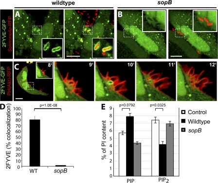
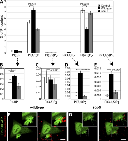
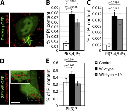
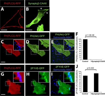
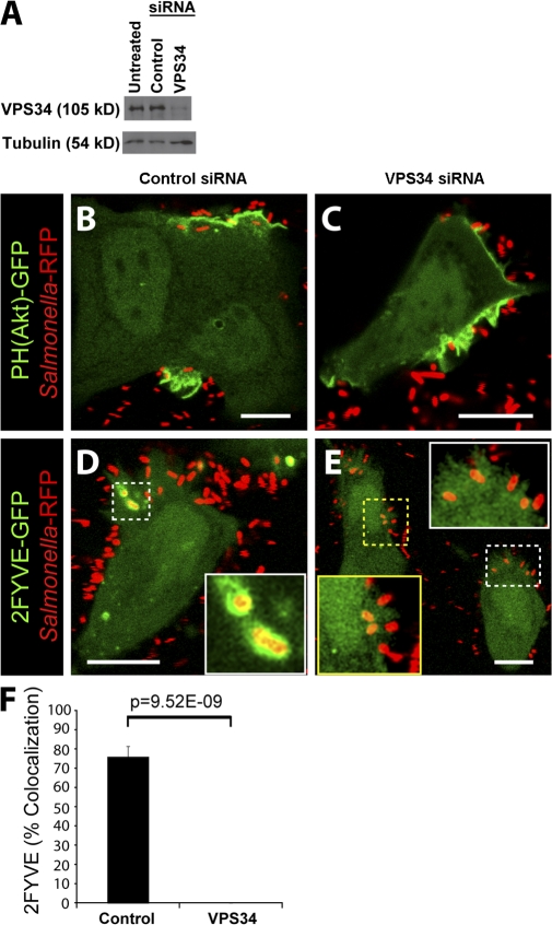
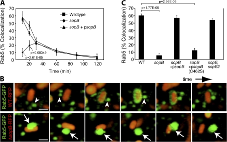
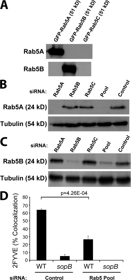
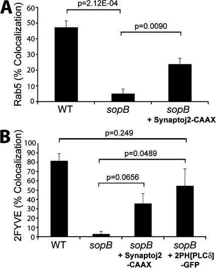
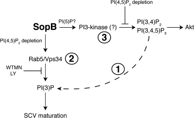
Salmonella colonizes a vacuolar niche in host cells during infection. Maturation of the Salmonella-containing vacuole (SCV) involves the formation of phosphatidylinositol 3-phosphate (PI(3)P) on its outer leaflet. SopB, a bacterial virulence factor with phosphoinositide phosphatase activity, was proposed to generate PI(3)P by dephosphorylating PI(3,4)P2, PI(3,5)P2, and PI(3,4,5)P3. Here, we examine the mechanism of PI(3)P formation during Salmonella infection. SopB is required to form PI(3,4)P2/PI(3,4,5)P3 at invasion ruffles and PI(3)P on nascent SCVs. However, we uncouple these events experimentally and reveal that SopB does not dephosphorylate PI(3,4)P2/PI(3,4,5)P3 to produce PI(3)P. Instead, the phosphatase activity of SopB is required for Rab5 recruitment to the SCV. Vps34, a PI3-kinase that associates with active Rab5, is responsible for PI(3)P formation on SCVs. Therefore, SopB mediates PI(3)P production on the SCV indirectly through recruitment of Rab5 and its effector Vps34. These findings reveal a link between phosphoinositide phosphatase activity and the recruitment of Rab5 to phagosomes.


Figures

Figure 1.
Figure 1.
SopB is required for PI(3)P localization to SCVs and an elevation of cellular PIP. (A and B) Cells were transfected with 2FYVE-GFP and infected with WT S. typhimurium (A) or a sopb deletion mutant (B). Left panels indicate 2FYVE-GFP relative to the RFP-expressing bacteria (red) in the merged images in the right panels. Insets are enlarged from the indicated areas (dashed boxes). Cells were analyzed using confocal microscopy. These images correspond to <15 min of infection. (C) Cells were cotransfected with PH(Akt)-RFP and 2FYVE-GFP before infection by WT S. typhimurium. Images were acquired at 1-min intervals. The time after infection of each frame is indicated. Insets are enlarged from the dashed boxes. Bars, 10 μm. (D) Cells were transfected with 2FYVE-GFP, infected with WT or sopB mutant, and analyzed as in A and B. Images were acquired at 1-min intervals for at least 1 h. Colocalization of the RFP-expressing bacteria with 2FYVE-GFP during the course of infection is shown. Data are means ± SEM of four separate experiments for WT bacteria (137 SCVs analyzed) and three separate experiments for sopB mutant (76 SCVs analyzed). (E) Phosphoinositide levels were quantified before (control) and after infection with either WT or sopB mutant. Lipids were extracted and analyzed by HPLC. The amount of the phosphoinositides is given as the percentage of PI in the same sample. PIP refers to the sum of PIPs. PIP2 refers to the sum of PIP2s. Data are means ± SEM of five separate experiments. The p-values are shown.
Figure 2.
Figure 2.
SopB mediates PI3-kinase activation at the plasma membrane during Salmonella invasion. (A) Lipids were extracted and analyzed by HPLC. The amount of phosphoinositides is given as the percentage of PI in the same sample. PI(4/5)P refers to the sum of PI(4)P and PI(5)P. (B–E) Insets are provided (arrows) for each 3′-phosphorylated phosphoinositide. Data are means ± SEM of five separate experiments. Levels of significance are indicated by p-values. (F and G) Cells were transfected with PH(Akt)-GFP and infected with WT (F) or sopB mutant (G). Cells were analyzed using confocal microscopy. Left panels indicate the PH(Akt)-GFP localization relative to the RFP- expressing bacteria (red) in the merged images in the right panels. Insets are enlarged from dashed boxes. These images correspond to <15 min of infection. Bars, 10 μm.
Figure 3.
Figure 3.
PI(3)P formation on the SCV is sensitive to pharmacologic inhibitors of PI3-kinase, but PI(3,4)P2 and PI(3,4,5)P3 production at invasion ruffles is not. (A) Cells were transfected with PH(Akt)-GFP and infected with WT S. typhimurium in the presence of LY294002. (B, C, and E) Phosphoinositide levels were quantified by HPLC in control cells and after infection with WT in the absence or presence of LY294002, as indicated. The amount of PI(3,4)P2 (B), PI(3,4,5)P3 (C), and PI(3)P (E) is given as the percentage of PI in the same sample. (D) Cells were transfected with 2FYVE-GFP and infected with WT in the presence of LY294002. The inset is enlarged from the indicated area (dashed box). Data in B, C, and E are means ± SEM of five separate experiments. Levels of significance are indicated by p- values. Cells in A and D were analyzed by confocal microscopy and correspond to <15 min of infection. Bars, 10 μm.
Figure 4.
Figure 4.
PI(3)P formation on the SCV is not dependent on PI(3,4)P2 and PI(3,4,5)P3 production at invasion ruffles. (A and B) Localization of PH(PLCδ)-RFP (A) and synaptojanin-2–CAAX construct (B) in control cells. Arrowheads indicate plasma membrane localization of synaptojanin-2–CAAX construct. (C–F) Cells were cotransfected with synaptojanin-2–CAAX, PH(PLCδ)-RFP (C), and PH(Akt)-GFP (D). (E) Merged image showing localization of bacteria (labeled with Alexa Fluor 647) relative to signal for PH(Akt)-GFP. (F) Cells were cotransfected as in C–E, infected with WT S. typhimurium, and analyzed by confocal microscopy. Images were acquired at 1-min intervals for ∼1 h. Colocalization of the bacteria with PH(Akt)-GFP during infection is shown. As a control, cells were cotransfected with PH(PLCδ)-RFP and PH(Akt)-GFP but not synaptojanin-2–CAAX. Data are means ± SEM of three separate experiments for synaptojanin-2–CAAX-expressing cells (20 ruffles analyzed) and two separate experiments for control cells (13 ruffles analyzed). The p-value is shown. (G–J) Cells were cotransfected with synaptojanin-2–CAAX, PH(PLCδ)-RFP (G), and 2FYVE-GFP (H). (I) Merged image showing localization of bacteria (labeled with Alexa Fluor 647) relative to signal for 2FYVE-GFP. Insets are enlarged from dashed boxes. (J) Cells were cotransfected as in G–I, infected with WT bacteria, and analyzed by confocal microscopy. Images were acquired at 1-min intervals for at least 1 h. Colocalization of the bacteria with 2FYVE-GFP during infection is shown. As a control, cells were cotransfected with PH(PLCδ)-RFP and 2FYVE-GFP but not synaptojanin-2–CAAX. Data are means ± SEM of three separate experiments for synaptojanin-2–CAAX- expressing cells (111 SCVs analyzed) and two separate experiments for control cells (44 SCVs analyzed). The p-value is shown. Bars, 10 μm.
Figure 5.
Figure 5.
Vps34 expression is required for PI(3)P formation on the SCV. (A) Western blot analysis of HeLa cell lysates. Tubulin expression was assessed as a loading control. (B–E) Cells were treated with control (B and D) or Vps34 (C and E) siRNA before transfection with either PH(Akt)-GFP (B and C) or 2FYVE-GFP (D and E). Cells were then infected with WT bacteria (Salmonella-RFP) and analyzed by confocal microscopy. Insets in D and E are enlarged from dashed boxes. These images correspond to <15 min of infection. Bars, 10 μm. (F) Cells were treated with control or Vps34 siRNA and infected with WT bacteria expressing RFP. Infected cells were analyzed using confocal microscopy. Images were acquired at 1-min intervals for ∼1 h. Colocalization of the bacteria with 2FYVE-GFP during infection is shown. Data are means ± SEM of four separate experiments for control siRNA–treated cells (102 SCVs analyzed) and three separate experiments for Vps34 siRNA–treated cells (170 SCVs analyzed). The p-value is shown.
Figure 6.
Figure 6.
Rab5 recruitment to the SCV requires the phosphatase activity of SopB. (A) Cells expressing Rab5A-GFP were infected for designated times and were fixed and stained for bacteria. Colocalization of Rab5A-GFP with intracellular bacteria was quantified. Data are means ± SEM of three separate experiments. Levels of significance are indicated by p-values compared with WT-infected cells. (B) Cells were transfected with Rab5A-GFP and infected with WT bacteria (WT-RFP, top) or an sopB mutant expressing RFP (sopB-RFP, bottom). Live imaging was performed on a spinning disk confocal microscope. Total sequence time for both series is ∼3 min. Arrowheads indicate Rab5A+ endosomes that dock and fuse with WT SCVs. Arrows indicate Rab5A+ endosomes that dock but do not fuse with sopB SCVs. Bar, 2.5 μm. (C) Cells were transfected with Rab5A-GFP and infected with either WT or sopB-deficient bacteria. In parallel, cells were infected with sopB mutant bacteria expressing either WT sopB (sopB + psopB) or a catalytically inactive mutant (sopB + psopB[C462S]) on a plasmid. A mutant lacking sopE and sopE2 was also analyzed. At 10-min after infection, infected cells were fixed and stained as in A. Colocalization of Rab5A-GFP with intracellular bacteria was quantified. Data are means ± SEM of three separate experiments. The p-values are shown.
Figure 7.
Figure 7.
Rab5 expression is required for PI(3)P localization to the SCV. (A) Cells were transfected with GFP-tagged Rab5A, Rab5B, or Rab5C as indicated. Cell lysates were then immunoblotted for either Rab5A or Rab5B. (B) Cells were treated with siRNA to the indicated Rab5 isoform, all three Rab5 isoforms (Pool), or control siRNA that does not affect Rab5 expression. Cell lysates were immunoblotted for Rab5A or tubulin (loading control). (C) Samples from B were immunoblotted for Rab5B or tubulin. (D) Cells were treated with either control siRNA or Rab5 Pool siRNA and transfected with 2FYVE-GFP. Cells were then infected with either WT or sopb mutant bacteria, fixed at 10-min after infection, and stained for bacteria. Intracellular bacteria colocalizing with 2FYVE-GFP was then quantified. Data are means ± SEM of three separate experiments (>100 bacteria analyzed per experiment). The p-value is shown.
Figure 8.
Figure 8.
Depletion of PI(4,5)P2 partially complements Rab5 and PI(3)P localization to sopB mutant SCVs. (A) Cells were cotransfected with Rab5A-GFP and PH(PLCδ)-RFP or synaptojanin-2–CAAX, PH(PLCδ)-RFP, and Rab5A-GFP as indicated. Cells were then infected with either WT or sopB mutant expressing RFP, fixed at 10-min after infection, and stained for extracellular bacteria. Intracellular bacteria colocalizing with Rab5A-GFP was then determined by microscopic analysis. Data are means ± SEM of three separate experiments (>100 bacteria analyzed per experiment). (B) Cells were transfected with the indicated construct and then infected with either WT or sopB mutant labeled covalently with Alexa Fluor 647. Infected cells were analyzed using a spinning disk confocal microscope. Images were acquired at 1-min intervals for ∼1 h. Localization of PI(3)P to SCVs (colocalization with 2FYVE-GFP or PX-mCherry) during the course of infection is shown. Data are means ± SEM of three separate experiments for WT- (94 SCVs analyzed) or sopB mutant– (68 SCVs analyzed) infected cells expressing 2FYVE-GFP. Five separate experiments were analyzed for sopb mutant–infected cells expressing synaptojanin 2-CAAX, PH(PLCδ)-RFP, and 2FYVE-GFP (121 SCVs analyzed), and three separate experiments were analyzed for sopb mutant–infected cells expressing 2PH(PLCδ)-GFP and PX-mCherry-GFP (120 SCVs analyzed). The p-values are shown.
Figure 9.
Figure 9.
Model of SopB action in host cells during Salmonella infection. During invasion, S. typhimurium injects SopB into host cells. At the plasma membrane, SopB mediates dephosphorylation of PI(4,5)P2 to generate PI(4/5)P. Through this event, SopB causes the accumulation of PI(3,4)P2 and PI(3,4,5)P3 at invasion ruffles by a wortmannin/LY294002-insensitive mechanism. SopB-mediated dephosphorylation of PI(4,5)P2 also allows SCV fission from the plasma membrane and the recruitment of Rab5 to nascent SCVs via their fusion with early endosomes. The Rab5 effector Vps34 catalyzes phosphorylation of PI to generate PI(3)P on SCVs, leading to downstream SCV maturation events such as the recruitment of LAMP-1.

References

    1. Anderson, K.E., and S.P. Jackson. 2003. Class I phosphoinositide 3-kinases. Int. J. Biochem. Cell Biol. 35:1028–1033. - PubMed
    1. Baldeon, M.E., B.P. Ceresa, and J.E. Casanova. 2001. Expression of constitutively active Rab5 uncouples maturation of the Salmonella-containing vacuole from intracellular replication. Cell. Microbiol. 3:473–486. - PubMed
    1. Birmingham, C.L., A.C. Smith, M.A. Bakowski, T. Yoshimori, and J.H. Brumell. 2006. Autophagy controls Salmonella infection in response to damage to the Salmonella-containing vacuole. J. Biol. Chem. 281:11374–11383. - PubMed
    1. Brumell, J.H., and S. Grinstein. 2004. Salmonella redirects phagosomal maturation. Curr. Opin. Microbiol. 7:78–84. - PubMed
    1. Bucci, C., A. Lutcke, O. Steele-Mortimer, V.M. Olkkonen, P. Dupree, M. Chiariello, C.B. Bruni, K. Simons, and M. Zerial. 1995. Co-operative regulation of endocytosis by three Rab5 isoforms. FEBS Lett. 366:65–71. - PubMed

Publication types

MeSH terms

Substances