Review
. 2008 Sep 1;112(5):1570-80.
doi: 10.1182/blood-2008-02-078071.

B lymphocytes: how they develop and function

Affiliations
Review

B lymphocytes: how they develop and function

Tucker W LeBien et al. Blood. .

Abstract

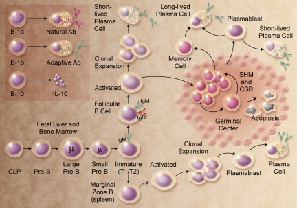
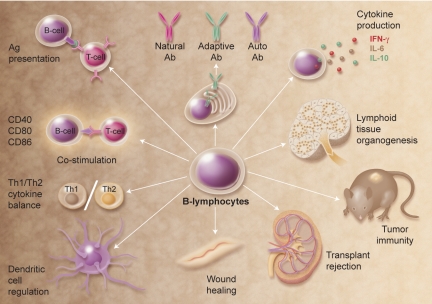
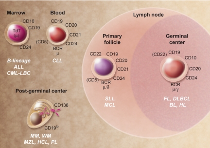
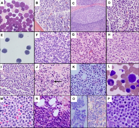
The discovery that lymphocyte subpopulations participate in distinct components of the immune response focused attention onto the origins and function of lymphocytes more than 40 years ago. Studies in the 1960s and 1970s demonstrated that B and T lymphocytes were responsible primarily for the basic functions of antibody production and cell-mediated immune responses, respectively. The decades that followed have witnessed a continuum of unfolding complexities in B-cell development, subsets, and function that could not have been predicted. Some of the landmark discoveries that led to our current understanding of B lymphocytes as the source of protective innate and adaptive antibodies are highlighted in this essay. The phenotypic and functional diversity of B lymphocytes, their regulatory roles independent of antibody production, and the molecular events that make this lineage unique are also considered. Finally, perturbations in B-cell development that give rise to certain types of congenital immunodeficiency, leukemia/lymphoma, and autoimmune disease are discussed in the context of normal B-cell development and selection. Despite the significant advances that have been made at the cellular and molecular levels, there is much more to learn, and cross-disciplinary studies in hematology and immunology will continue to pave the way for new discoveries.


Figures

None
Thomas F. Tedder
None
Tucker W. LeBien
Figure 1
Figure 1
B-cell development. The figure shows the broad outline of B-cell developmental stages in mice and humans. B-1a, B-1b, and B10 populations are less well characterized in humans. CLP indicates common lymphoid progenitor; SHM, somatic hypermutation; and CSR, class switch recombination. Illustration by A. Y. Chen.
Figure 2
Figure 2
Multifunctional attributes of B cells. Selected examples of how B cells regulate immune homeostasis are shown; many of these functions are independent of Ig production. Illustration by A. Y. Chen.
Figure 3
Figure 3
Human B-cell malignancies. Selected cell surface, cytoplasmic, and nuclear markers expressed during normal B-cell development that are generally expressed in malignancies. Molecules within parentheses are variable in their expression at the indicated stage of development. ALL indicates acute lymphoblastic leukemia; BL, Burkitt lymphoma; CLL, chronic lymphocytic leukemia; CML-LBC, chronic myelocytic leukemia–lymphoid blast crisis; DLBCL, diffuse large B-cell lymphoma; FL, follicular lymphoma; HCL, hairy cell leukemia; HL, Hodgkin lymphoma; MCL, mantle cell lymphoma; MM, multiple myeloma; MZL, marginal zone lymphoma; PL, plasmablastic lymphoma; SLL, small lymphocytic lymphoma; and WM, Waldenström macroglobulinemia. Illustration by A. Y. Chen.
Figure 4
Figure 4
A histopathological montage of normal and malignant human B-lineage cells. (A) Bone marrow aspirate of ALL cells exhibiting a high nuclear to cytoplasmic ratio, finely dispersed chromatin, indistinct nucleoli, and blue-gray cytoplasm. (B) Bone marrow biopsy showing effacement of the marrow architecture by a monotonous population of ALL lymphoblasts. (C) Representative normal lymph node follicle showing a reactive GC surrounded by mantle zone. (D) MCL cells display a heterogeneous morphology ranging from small round (true mantle zone/CLL-like) cells to those showing irregular cleaved nuclei (FL-like). (E) A peripheral blood sample of CLL cells exhibiting closely condensed chromatin, indistinct nucleoli, and scant basophilic cytoplasm with a regular outline. (F) Normal GC centrocytes with vesicular chromatin and irregular nuclear outlines, along with larger round centroblasts showing a similar open chromatin. (G) FL with a predominance of small cleaved centrocyte-like cells demonstrating significant nuclear irregularity. (H) DLBCL with classic post-GC immunoblastic morphology exhibiting a large nucleus, eosinophilic nucleoli, vesicular chromatin, and relatively abundant cytoplasm. (I) Biopsy from an abdominal mass showing BL cells with a monomorphic infiltrate of small to intermediate-sized cells with round nuclei, indistinct nucleoli, clumped chromatin, a basophilic cytoplasm, and a high apoptotic cell index. (J) A characteristic Reed-Sternberg cell (arrow) in HL. The cells are large and binucleated, with mirror image nuclei, demonstrating prominent eosinophilic nucleoli. Many of the other cells in the background are reactive lymphocytes. (K) Bone marrow biopsy showing hairy cell leukemia. The cells are evenly spaced from each other with round nuclei and clear abundant cytoplasm, imparting a “fried egg” appearance to the cells. (L) Peripheral blood smear of hairy cell leukemia (HCL) demonstrating classic concentric hairlike cytoplasmic projections. (M) MZL cells with a heterogeneous appearance ranging from small round cells to those manifesting a cleaved morphology. (N) WM/lymphoplasmacytic lymphoma showing considerable morphologic overlap with MZL. The cells demonstrate significant plasmacytoid features with pseudointranuclear inclusions (ie, Dutcher bodies). Many of the plasmacytoid cells contain globular collections of paraprotein in the cytoplasm, imparting a mulberry-like appearance to the cell. (O) Two cases of MM within the marrow. The cells show typical plasma cell features including the eccentrically disposed nucleus and abundant Ig-containing cytoplasm. (P) PL comprising enlarged plasmacytoid cells with enhanced atypia; mitoses are easily identifiable. All photographs were taken with an Olympus BX41 microscope and DP71 camera (Olympus America, Center Valley, PA). The images were acquired using the following UPlanFL N objective lenses: panels A,E,J,L,M,O, P: 100×/1.30, oil immersion; panels B,F,G,H,I,K,N: 40×/0.75; panels C,D: 20×/0.50. Panels A, L, and O are Giemsa stains; all others are H and E stains. Images were assembled using Adobe Photoshop version 9.0.2 (Adobe Systems, San Jose, CA). Panels A and E were provided by Dr Amy Chadburn, and all other images were provided by Dr Cynthia Magro, both from the Weill Medical College of Cornell University, New York, NY.

References

    1. Cooper MD, Alder MN. The evolution of adaptive immune systems. Cell. 2006;124:815–822. - PubMed
    1. Tiselius A, Kabat EA. Electrophoresis of immune serum. Science. 1938;87:416–417. - PubMed
    1. Fagraeus A. The plasma cellular reaction and its relation to the formation of antibodies in vitro. J Immunol. 1948;58:1–13. - PubMed
    1. Jerne NK. The natural-selection theory of antibody formation. Proc Natl Acad Sci U S A. 1955;41:849–857. - PMC - PubMed
    1. Burnet FM. A modification of Jerne's theory of antibody production using the concept of clonal selection. Aust J Sci. 1957;20:67–69. - PubMed

MeSH terms

Substances