. 2008 Sep 9;105(36):13409-14.
doi: 10.1073/pnas.0806136105. Epub 2008 Aug 25.

Heparin promotes the growth of human embryonic stem cells in a defined serum-free medium

Affiliations

Heparin promotes the growth of human embryonic stem cells in a defined serum-free medium

Miho K Furue et al. Proc Natl Acad Sci U S A. .

Erratum in

  • Proc Natl Acad Sci U S A. 2008 Nov 18;105(46):18071

Abstract

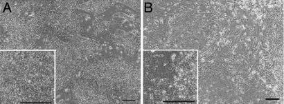
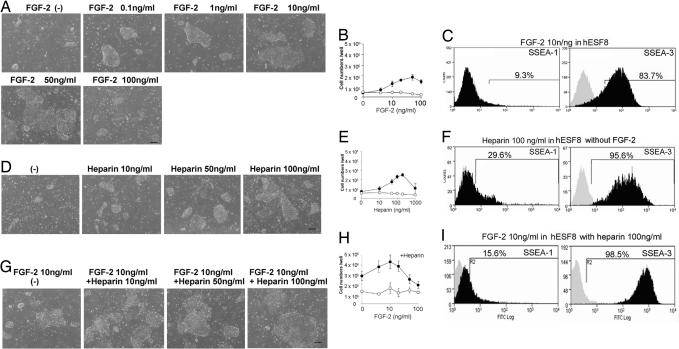
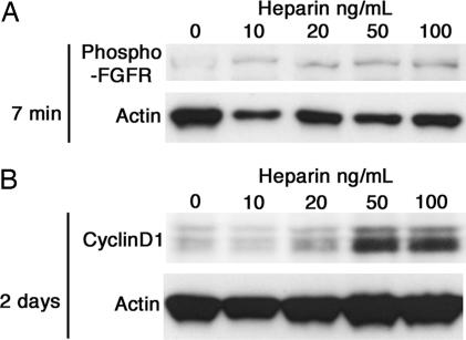
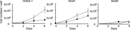
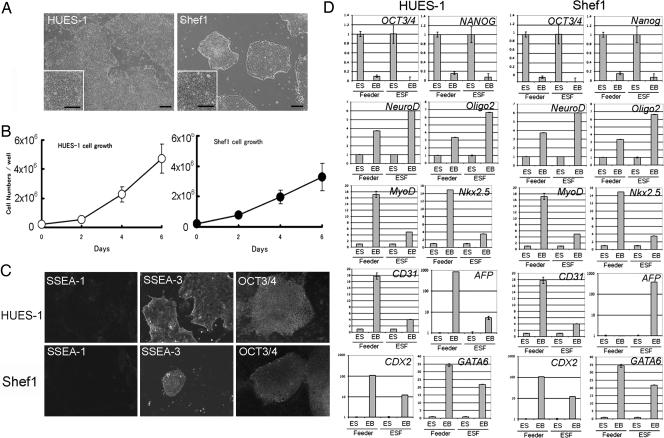
A major limitation in developing applications for the use of human embryonic stem cells (HESCs) is our lack of knowledge of their responses to specific cues that control self-renewal, differentiation, and lineage selection. HESCs are most commonly maintained on inactivated mouse embryonic fibroblast feeders in medium supplemented with FCS, or proprietary replacements such as knockout serum-replacement together with FGF-2. These undefined culture conditions hamper analysis of the mechanisms that control HESC behavior. We have now developed a defined serum-free medium, hESF9, for the culture of HESCs on a type I-collagen substrate without feeders. In contrast to other reported media for the culture of HESCs, this medium has a lower osmolarity (292 mosmol/liter), l-ascorbic acid-2-phosphate (0.1 microg/ml), and heparin. Insulin, transferrin, albumin conjugated with oleic acid, and FGF-2 (10 ng/ml) were the only protein components. Further, we found that HESCs would proliferate in the absence of exogenous FGF-2 if heparin was also present. However, their growth was enhanced by the addition of FGF-2 up to 10 ng/ml although higher concentrations were deleterious in the presence of heparin.


Conflict of interest statement

The authors declare no conflict of interest.

Figures

Fig. 1.
Fig. 1.
Phase contrast photomicrograph of HUES-1 (A) and Shef 1 (B) HESCs cultured on feeders in KSR-based medium. (Scale bar, 200 μm.)
Fig. 2.
Fig. 2.
Effect of FGF-2 and heparin on the HUES-1 cell growth. HUES-1 cells were cultured on type I collagen in hESF8 with varying concentrations of FGF-2 or heparin. (A) Phase contrast photomicrograph of HUES-1 cells with various concentration of FGF-2 without heparin. (Scale bar, 200 μm.) (B) Number cells harvested after 6 days of culture in KSR-based medium (open circles) or hESF8 (closed circles). Values are the mean ± SD for three measurements. The absence of error bars is due to the small SD in the results. (C) FACS profiles for SSEA-1 and SSEA-3 expression of cells cultured with 10 ng/ml FGF-2. Antigen histogram (black); control histogram (gray); the horizontal bar indicates the gating used to score the percentage of cells antigen positive. Similar profiles were obtained for cells from cultures grown with all concentrations of FGF-2 from 0 to 100 ng/ml (data not shown). (D) Phase contrast photomicrograph of HUES-1 cells cultured without FGF-2 but supplemented with various concentration of heparin. (Scale bar, 200 μm.) (E) Number cells harvested after 6 days of culture in KSR-based medium (open circles) or hESF8 (closed circles) with varying concentrations of heparin but no FGF-2. Values are the mean ± SD for three measurements. The absence of error bars is due to the small SD in the results. (F) FACS profiles for SSEA-1 and SSEA-3 expression of cells cultured with 100 ng/ml heparin. Antigen histogram (black); control histogram (gray); the horizontal bar indicates the gating used to score the percentage of cells antigen positive. Similar profiles were obtained for cells from cultures grown with all concentrations of heparin from 0 to 100 ng/ml (data not shown). (G) Phase contrast photomicrograph of HUES-1 cells cultured with various concentration of heparin in hESF8 containing 10 ng/ml FGF-2. (Scale bar, 200 μm.) (H) Number cells harvested after 6 days of culture in KSR-based medium (open circles) or hESF8 (closed circles) containing varying concentrations of FGF-2 and 100 ng/ml heparin. Values are the mean ± SD for three measurements. The absence of error bars is due to the small SD in the results. (I) FACS profiles for SSEA-1 and SSEA-3 expression of cells cultured hESF8 containing 100 ng/ml heparin and 10 ng/ml FGF-2 (hESF9). Antigen histogram (black); control histogram (gray); the horizontal bar indicates the gating used to score the percentage of antigen-positive cells. Similar profiles were obtained for cells from cultures grown with all concentrations of FGF-2 from 0 to 100 ng/ml (data not shown).
Fig. 3.
Fig. 3.
The effect of heparin on FGF signaling. HUES-1 cells were stimulated with different concentrations of heparin after 48-h starvation of FGF-2 and heparin. (A) Seven minutes after heparin addition, the cells were lysed and followed by western blot using an antibody detecting the phosphorylation of FGF receptors. (B) After 2 days of culture in the presence of heparin, the cells were lysed and subjected to western blot of cyclin D1. Actin is used as loading control.
Fig. 4.
Fig. 4.
Effect of ECM on HUES-1 culture. (A–E) Phase contrast photomicrographs of HUES-1 cells cultured in hESF9 medium, in flasks coated with collagen (A), gelatin (B), fibronectin (C), laminin (D), and Matrigel (E). (F) Cells were cultured in KSR-based medium on type I collagen. (Scale bar, 200 μm.)
Fig. 5.
Fig. 5.
FACS profiles for SSEA-1, SSEA-3, and TRA-1–60 expression by HUES-1, Shef1, Shef4, Shef5, and H7 HESC lines cultured on type I collagen in hESF9, in comparison with cells grown on feeders in KSR-based medium. Antigen histogram (black); control histogram (gray); the horizontal bar indicates the gating used to score the percentage of antigen-positive cells.
Fig. 6.
Fig. 6.
A comparison of the growth of different HESCs in hESF9 and KSR-based media. HUES-1, Shef1, and Shef5 cells were seeded on feeders in KSR-based medium (closed circles) or on type I collagen in hESF9 (open circles) at a cell density of 1 × 10⁵ cells per well; mean and SD of three experiments. Cell numbers were counted every 2 days.
Fig. 7.
Fig. 7.
Long-term culture of HESCs in the defined medium. HUES-1 and Shef1 cells were serially cultured on type I collagen in hESF9. The cells were split at 1:3 every week. (A) Phase contrast photomicrograph of HUES-1 at passage 21 and Shef1 cells at passage 14. (Scale bar, 200 μm.) (B) The growth of HUES-1 (passage 10) and Shef1 cells (passage 5) in the defined medium. (C) Immunohistochemical staining of HUES-1 (passage 24) and Shef1 (passage 14) for SSEA-1, SSEA-3, and OCT3/4. (D) Q-PCR analysis of gene expression in HUES-1 and Shef1 on feeder in KSR-based medium (feeder) and HUES-1 (passage 24) and Shef1 (passage 14) on collagen in hESF9 medium (ESF) those during in vitro differentiation (EB). The name of the gene of differentiation are noted in each bar graph. Expression levels were all normalized against GAPDH. The relative level of each gene in undifferentiated cells was defined as “1.”

References

    1. Thomson JA, et al. Embryonic stem cell lines derived from human blastocysts. Science. 1998;282:1145–1147. - PubMed
    1. Reubinoff BE, Pera MF, Fong CY, Trounson A, Bongso A. Embryonic stem cell lines from human blastocysts: Somatic differentiation in vitro. Nat Biotechnol. 2000;18:399–404. - PubMed
    1. Xu C, et al. Feeder-free growth of undifferentiated human embryonic stem cells. Nat Biotechnol. 2001;19:971–974. - PubMed
    1. Smith AG, et al. Inhibition of pluripotential embryonic stem cell differentiation by purified polypeptides. Nature. 1988;336:688–690. - PubMed
    1. Williams RL, et al. Myeloid leukaemia inhibitory factor maintains the developmental potential of embryonic stem cells. Nature. 1988;336:684–687. - PubMed

Publication types