. 2008 Sep 2;105(35):13093-8.
doi: 10.1073/pnas.0805213105. Epub 2008 Aug 25.

Structural basis for suppression of a host antiviral response by influenza A virus

Affiliations

Structural basis for suppression of a host antiviral response by influenza A virus

Kalyan Das et al. Proc Natl Acad Sci U S A. .

Abstract

Influenza A viruses are responsible for seasonal epidemics and high mortality pandemics. A major function of the viral NS1A protein, a virulence factor, is the inhibition of the production of IFN-beta mRNA and other antiviral mRNAs. The NS1A protein of the human influenza A/Udorn/72 (Ud) virus inhibits the production of these antiviral mRNAs by binding the cellular 30-kDa subunit of the cleavage and polyadenylation specificity factor (CPSF30), which is required for the 3' end processing of all cellular pre-mRNAs. Here we report the 1.95-A resolution X-ray crystal structure of the complex formed between the second and third zinc finger domain (F2F3) of CPSF30 and the C-terminal domain of the Ud NS1A protein. The complex is a tetramer, in which each of two F2F3 molecules wraps around two NS1A effector domains that interact with each other head-to-head. This structure identifies a CPSF30 binding pocket on NS1A comprised of amino acid residues that are highly conserved among human influenza A viruses. Single amino acid changes within this binding pocket eliminate CPSF30 binding, and a recombinant Ud virus expressing an NS1A protein with such a substitution is attenuated and does not inhibit IFN-beta pre-mRNA processing. This binding pocket is a potential target for antiviral drug development. The crystal structure also reveals that two amino acids outside of this pocket, F103 and M106, which are highly conserved (>99%) among influenza A viruses isolated from humans, participate in key hydrophobic interactions with F2F3 that stabilize the complex.


Conflict of interest statement

The authors declare no conflict of interest.

Figures

Fig. 1.
Fig. 1.
Crystal structure of F2F3:NS1A (85-215) complex. (A) Gel filtration data demonstrating complex formation between NS1A (85-215) and F2F3. Traces show chromatographic profiles on a Superdex G75 column for NS1A (85-215) alone (red) and the complex of NS1A (85-215) with [S94]-F2F3 (blue). Inset shows calibrated gel filtration data for (A) [S94]-F2F3 (∼10 kDa), (B) NS1A (85-215) alone (∼27 kDa), and (C) [S94]-F2F3:NS1A complex (∼47 kDa). Similar results were obtained by static light scattering analysis of effluent fractions from size exclusion chromatography, as described in SI Materials and Methods: (A) 15 ± 3 kDa, (B) 25 ± 5 kDa, and (C) 48 ± 5 kDa. The molecular mass expected for the tetrameric complex observed in the crystal structure is 49,250 Da, which is in good agreement with these light scattering and gel filtration data. The elution times for isolated NS1A (85-215) (single chain calculated molecular mass 15,943 Da) and [S94]-F2F3 (single chain calculated molecular mass 8,682 Da) domains differ when loaded at different protein concentrations, suggesting that these molecules form weak homodimers under these solution conditions. Calibration standards (A–D) are described in SI Materials and Methods. (B) Two NS1A effector domains (green and red) and two F2F3 domains (blue and yellow) of CPSF30 form the tetramer. Some NS1A′ amino acid residues that function in complex formation are highlighted in cyan. (C) F3-binding pocket on NS1A (85-215). A hydrophobic pocket on the NS1A surface binds to the F3 Zn finger of F2F3. Both chains of NS1A in the head-to-head dimer interact with each F2F3 molecule. (D) Expanded view of the F3-binding pocket. The NS1A amino acid residues labeled in red interact with the aromatic side chains of residues Y97, F98, and F102 of the F3 Zn finger of F2F3.
Fig. 2.
Fig. 2.
Effects of amino acid substitutions in the NS1A protein on its interaction with CPSF30 and on its function in influenza A virus-infected cells. (A) GST-pulldown assay. GST-F2F3 or GST were mixed with equal amounts of the WT or indicated mutant 35S-labeled full-length NS1A protein of Ud, which were prepared as described in SI Materials and Methods. The labeled proteins eluted with glutathione from GST-F2F3 or GST were resolved by SDS-polyacrylamide gels, which were analyzed by exposure to X-ray film. (B) Plaque sizes of the WT and G184R mutant Ud viruses in Madin-Darby canine kidney (MDCK) cells. (C) The G184R mutation in the Ud NS1A protein does not affect the amount of the NS1A protein synthesized in MDCK cells infected with 5 pfu/cell. Immunoblots of cell extracts collected at 6 h after infection were carried out with either anti-NS1A or antitubulin antibody. (D) Quantitative RT-PCR measuring amounts of IFN-β pre-mRNA (Left) and IFN-β mRNA (Right) in WT and G184R Ud-infected cells. Pre-mRNA results were normalized to WT, and mRNA results were normalized to G184R data. The results show the average and standard deviation for the relative levels of G184R pre-mRNA and WT mRNA from three different virus infections.
Fig. 3.
Fig. 3.
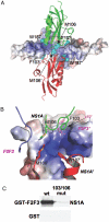
Structural role of F103 and M106 in formation of the tetrameric complex. (A) Molecular graphics showing how two F2F3 molecules (represented as the electrostatic potential surface) wrap around two NS1A (85-215) molecules. The head-to-head interaction of NS1A molecules forms a docking surface for F2F3 binding. The side chain of residue M106 is critically positioned at the tetrameric epicenter and interacts with the other three molecules. (B) Expanded regions showing the structural environments of amino acid residues F103 and M106 of NS1A. The aromatic side chain of NS1A F103 interacts primarily with the hydrophobic amino acid residues L72′, Y88′ and P111′ that are present on the surface of F2F3′. (C) GST-pulldown assay. GST-F2F3 or GST was mixed with the WT or 103/106 mutant [F103L, M106I]-NS1A protein, and analyzed as described in the legend of Fig. 2A.
Fig. 4.
Fig. 4.
Molecular graphic showing the locations of NS1A residues F103 and M106 with respect to the F3-binding pocket. The tetrameric interface extends beyond the hydrophobic pocket of the NS1A effector domain (orange) which binds one F2F3 molecule (blue). Residues M106 and F103 (green surfaces) of the same NS1A effector domain interact with both the F2F3 (blue) and F2F3′ (yellow) molecules. The M106 sidechain of this NS1A effector domain also interacts with the M106′ side chain of the second NS1A′ molecule (red) at the tetrameric epicenter.
Fig. 5.
Fig. 5.
PR8 virus infection, unlike infection by Ud virus, does not activate IRF-3. HEL299 cells were either mock-infected (M, lane 1), infected with 5 pfu/cell of Ud virus (Ud, lane 2), infected with 5 pfu/cell of PR8 virus (PR8, lane 3), or infected with 5 pfu/cell of a recombinant Ud virus in which the Ud NS gene was replaced by the PR8 NS gene (Ud/NS-PR8, lane 4). At 7 h after infection, cell extracts were prepared, subjected to electrophoresis on a 7.5% native gel, and IRF-3 monomers and dimers were detected by Western immunoblotting using rabbit anti-IRF-3 antibody (36). An immunoblot with anti-NS1A antibody confirmed that equivalent amounts of the NS1A protein were synthesized in Ud, PR8, and Ud/NS-PR8 virus-infected cells (lanes 2–4). Further details are provided in the SI Materials and Methods.

References

    1. Hatada E, Takizawa T, Fukuda R. Specific binding of influenza A virus NS1 protein to the virus minus-sense RNA. in vitro J Gen Virol. 1992;73:17–25. - PubMed
    1. Wang W, et al. RNA binding by the novel helical domain of the influenza virus NS1 protein requires its dimer structure and a small number of specific basic amino acids. RNA. 1999;5:195–205. - PMC - PubMed
    1. Chien CY, et al. Biophysical characterization of the complex between double-stranded RNA and the N-terminal domain of the NS1 protein from influenza A virus: evidence for a novel RNA-binding mode. Biochemistry. 2004;43:1950–1962. - PubMed
    1. Min J, Krug RM. The primary function of RNA binding by the influenza A virus NS1 protein in infected cells: inhibiting the 2′-5′ OAS/RNase L pathway. Proc Natl Acad Sci USA. 2006;103:7100–7105. - PMC - PubMed
    1. Li S, Min JY, Krug RM, Sen GC. Binding of the influenza A virus NS1 protein to PKR mediates the inhibition of its activation by either PACT or double-stranded RNA. Virology. 2006;349:13–21. - PubMed

Publication types

Associated data

LinkOut - more resources