. 2008 Aug 26;3(8):e3063.
doi: 10.1371/journal.pone.0003063.

The critical role of partially exposed N-terminal valine residue in stabilizing GH10 xylanase from Bacillus sp.NG-27 under poly-extreme conditions

Affiliations

The critical role of partially exposed N-terminal valine residue in stabilizing GH10 xylanase from Bacillus sp.NG-27 under poly-extreme conditions

Amit Bhardwaj et al. PLoS One. .

Erratum in

  • PLoS ONE.2009;4(2). doi: 10.1371/annotation/aefbf6a4-298d-4173-907f-9c9a996249b2.. Bharadwaj, Amit [corrected to Bhardwaj, Amit]

Abstract

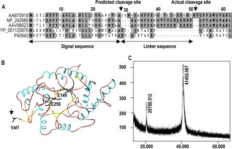
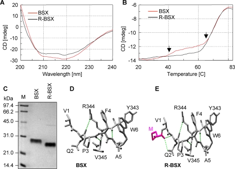
Background: Understanding the mechanisms that govern protein stability under poly-extreme conditions continues to be a major challenge. Xylanase (BSX) from Bacillus sp. NG-27, which has a TIM-barrel structure, shows optimum activity at high temperature and alkaline pH, and is resistant to denaturation by SDS and degradation by proteinase K. A comparative circular dichroism analysis was performed on native BSX and a recombinant BSX (R-BSX) with just one additional methionine resulting from the start codon. The results of this analysis revealed the role of the partially exposed N-terminus in the unfolding of BSX in response to an increase in temperature.

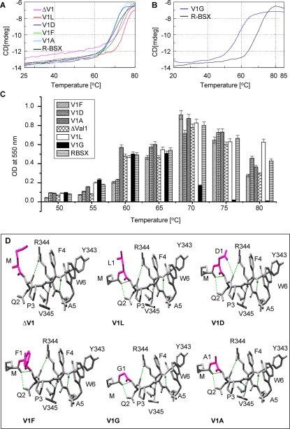
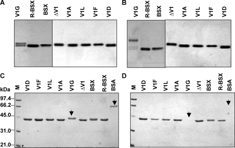
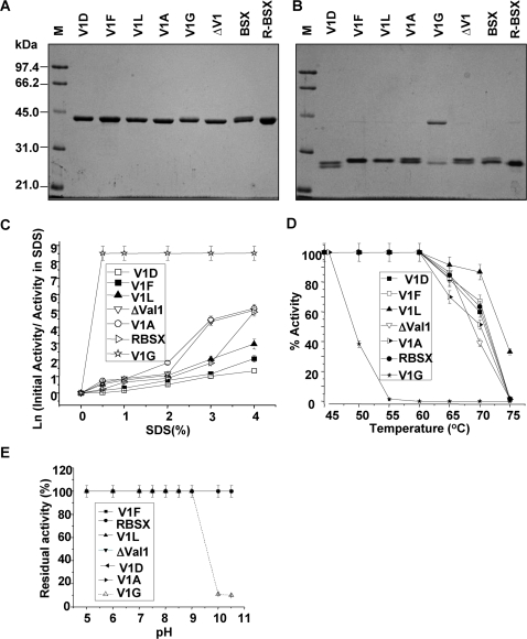
Methodology: We investigated the poly-extremophilicity of BSX to deduce the structural features responsible for its stability under one set of conditions, in order to gain information about its stability in other extreme conditions. To systematically address the role of the partially exposed N-terminus in BSX stability, a series of mutants was generated in which the first hydrophobic residue, valine (Val1), was either deleted or substituted with various amino acids. Each mutant was subsequently analyzed for its thermal, SDS and proteinase K stability in comparison to native BSX.

Conclusions: A single conversion of Val1 to glycine (Gly) changed R-BSX from being thermo- and alkali- stable and proteinase K and SDS resistant, to being thermolabile and proteinase K-, alkali- and SDS- sensitive. This result provided insight into the structure-function relationships of BSX under poly-extreme conditions. Molecular, biochemical and structural data revealed that the poly-extremophilicity of BSX is governed by a partially exposed N-terminus through hydrophobic interactions. Such hitherto unidentified N-terminal hydrophobic interactions may play a similar role in other proteins, especially those with TIM-barrel structures. The results of the present study are therefore of major significance for protein folding and protein engineering.


Conflict of interest statement

Competing Interests: The authors have declared that no competing interests exist.

Figures

Figure 1
Figure 1. Cleavage site of BSX signal sequence during extra-cellular secretion in Bacillus Sp. NG-27.
(A) Multiple sequence alignments of the BSX signal peptide with other GH10 xylanases of Bacillus origin. AAB70918: Bacillus sp. NG-27, used in the present study, NP_242986: Bacillus halodurans C-125, AAV98623: Bacillus halodurans S7, YP_001125870: Geobacillus thermodenitrificans NG80-2, P40943: Bacillus stearothermophilus T-6. (B) Ribbon diagram of BSX showing catalytic residues (E149 and E259) and the N-terminus Val1 residue (indicated by arrow). (C) Mass Spectrometry analysis of purified BSX confirming the processing of the first 51 amino acid-long signal sequence during its secretion instead of the predicted 28 amino acids. The predicted signal sequence and the additional linker sequence that is also processed are indicated by the double-headed arrows in (A).
Figure 2
Figure 2. Comparison of BSX and R-BSX structures using CD and SDS-PAGE.
(A) Far UV spectra of BSX and R-BSX. (B) CD spectra of thermal denaturation of BSX and R-BSX monitored at 222 nm with a temperature slope of 1°C/min. Note the difference in the thermal unfolding pattern between 40°C–60°C, marked by arrows. (C) SDS-PAGE analysis of BSX and R-BSX, showing higher mobility of R-BSX. To preserve their native conformations, samples were loaded onto the gel without prior boiling. (D) SwissPdb-generated structural models showing the N-terminal region of BSX and R-BSX. Additional hydrogen bonds between Met (start codon) and Gln2 are highlighted by colored dotted lines.
Figure 3
Figure 3. Thermal denaturation and structural features of the R-BSX mutants.
(A) CD spectra of thermal denaturation of ΔV1, V1L, V1D, V1F, V1A and R-BSX monitored at 222 nm with a temperature slope of 1°C/min. (B) CD spectra of thermal denaturation of V1G and R-BSX, showing the ∼12°C decrease in Tm for V1G. (C) Xylanase activity profile of R-BSX mutants at various temperatures. Maximum activity for V1G was observed at 60°C, differing from other mutants which showed maximum activity at 70°C similar to that of R-BSX. (D) SwissPdb-generated structural models showing the N-terminal region of all R-BSX variants. The amino acid substituted for Val1 is shown in pink. The additional hydrogen bonds resulting from a deletion/substitution are shown in green dotted lines.
Figure 4
Figure 4. Native PAGE profiles of BSX and R-BSX.
(A) and (B) show native-PAGE profiles of thermal unfolding of proteins at room temperature and at 60°C, respectively. V1G was found to unfold after incubation at 60°C for 15 min, whereas no significant unfolding was observed for all the other mutants. Note the slightly faster mobility of R-BSX when compared to BSX, reconfirming the results obtained in Figure 2C. (C) and (D) show SDS-PAGE profiles of all the proteins in the absence and presence of proteinase K, respectively. Note that all proteins except V1G and BSA (indicated by arrow) showed proteinase K resistance.
Figure 5
Figure 5. Effect of SDS and proteinase K on the BSX and R-BSX.
(A) and (B) show SDS-PAGE profiles for determining the SDS-stability of proteins. Identical protein samples were loaded onto the gel with or without prior boiling for 10 min, respectively. (C) Xylanase activity in the presence of SDS. Samples were incubated for 12 h in the presence of various concentrations of SDS prior to the activity assay. Note that V1F and V1D displayed maximum activity. (D) Thermostability of xylanase and its mutants. Samples were incubated for 15 min at various temperatures and assayed for residual xylanase activity at their respective optimum temperature (see material and methods). (E) Alkaline stability of BSX, R-BSX and its mutants. Note the drastic reduction in the activity of the V1G mutant when the enzyme was incubated above pH 10 prior to the assay.

References

    1. Alberti E, Consonni R, Zetta L. Applications of NMR to thermostable proteins. Annual Report in NMR Spectroscopy. 2003;50:121–161.
    1. Matthews BW. Structural and genetic analysis of protein stability. Annu Rev Biochem. 1993;62:139–160. - PubMed
    1. Fersht AR, Serrano L. Principles of protein stability derived from protein engineering experiments. Curr Opin Structuct Biol. 1993;3(1):75–83.
    1. Funahashi J, Tkano K, Yamagata Y, Yutani K. Role of surface hydrophobic residues in the conformational stability of human lysozyme at three diferent positions. Biochemistry. 2000;39(47):14448–14456. - PubMed
    1. Pakula AA, Sauer RT. Reverse hydrophobic effects relieved by amino-acid substitutions at a protein surface. Nature. 1990;344(6264):363–364. - PubMed

Publication types