. 2008 Dec 1;68(16):1773-89.
doi: 10.1002/pros.20845.

The dietary bioflavonoid, quercetin, selectively induces apoptosis of prostate cancer cells by down-regulating the expression of heat shock protein 90

Affiliations

The dietary bioflavonoid, quercetin, selectively induces apoptosis of prostate cancer cells by down-regulating the expression of heat shock protein 90

Ravikumar Aalinkeel et al. Prostate. .

Abstract

Background: Human and animal studies have suggested that diet-derived flavonoids, in particular quercetin may play a beneficial role by preventing or inhibiting oncogenesis, but the underlying mechanism remains unclear. The aim of this study is to evaluate the effect(s) of quercetin on normal and malignant prostate cells and to identify the target(s) of quercetin's action.

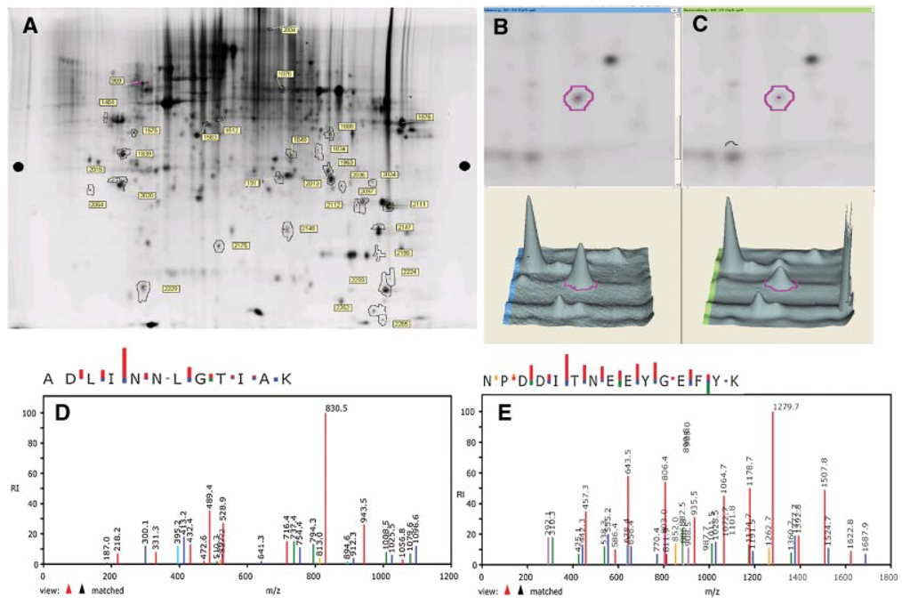
Methodology: We addressed this question using cells in culture and investigated whether quercetin affects key biological processes responsible for tumor cell properties such as cell proliferation and apoptosis and also studied the effect of quercetin on the proteome of prostate cancer cells using difference gel electrophoresis (DIGE) to assess changes in the expression of relevant proteins.

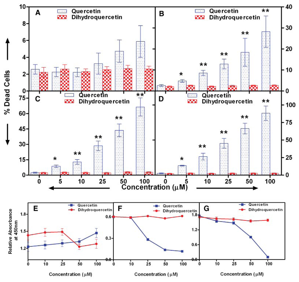
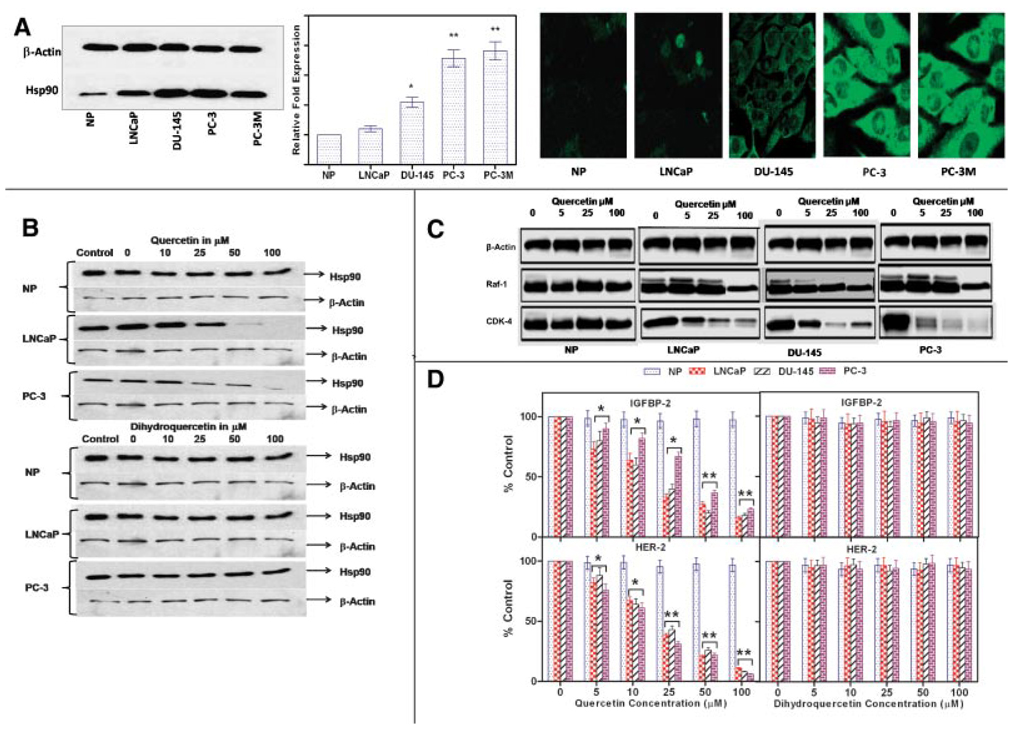
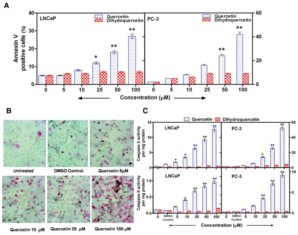
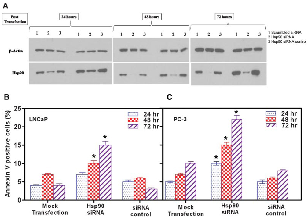
Results: Our findings demonstrate that quercetin treatment of prostate cancer cells results in decreased cell proliferation and viability. Furthermore, we demonstrate that quercetin promotes cancer cell apoptosis by down-regulating the levels of heat shock protein (Hsp) 90. Depletion of Hsp90 by quercetin results in decreased cell viability, levels of surrogate markers of Hsp90 inhibition (intracellular and secreted), induced apoptosis and activation of caspases in cancer cells but not in normal prostate epithelial cells. Knockdown of Hsp90 by short interfering RNA also resulted in induction apoptosis similar to quercetin in cancer cells as indicated by annexin V staining.

Conclusion: Our results demonstrate that quercetin down-regulates the expression of Hsp90 which, in turn, induces inhibition of growth and cell death in prostate cancer cells while exerting no quantifiable effect on normal prostate epithelial cells.


Figures

Fig. 1
Fig. 1
Effect of quercetin on the viability of prostate cancer cells. A: Normal prostate epithelial cells, (B) LNCaP, (C)DU-145, and (D) PC-3 prostate cancer cells were treated with increasing concentrations of quercetin for 24 hr and viability was determined by trypan blue vital dye exclusion. Viability was confirmed by a proprietary tetrazolium dye method using absorbance at 450 nm as described in the text: (E) normal prostate epithelial cells, (F) LNCaP, and (G) PC-3 cells. Values are mean ± SE of six experiments; *P < 0.05 and **P < 0.001 compared to treatment with dihydroquercetin which did not affect the viability of any of the cells in this study. [Color figure can be viewed in the online issue, which is available at http://www.interscience.wiley.com.]
Fig. 2
Fig. 2
Proteomic analysis on the effects of quercetin on prostate cancer cells showing that Hsp90 is maximally modulated (down-regulated) by treatment of PC-3 cells with quercetin. A: Representative 2D-Sypro-Ruby stained proteome image of quercetin treated PC-3 cells.PC-3 cells (1 × 10⁶) were cultured with 50µM of quercetin for 24hr. Total protein was extracted, subjected to DIGE analysis, and stained with SYPRO Ruby protein stain as described in the text. The pH increases from left to right and the molecular mass decreases from the top to the bottom of the gels. Identified protein spots are outlined and numbered, three separate experiments yielded similar results. Abundance of spot # 191 from (B) untreated and (C) quercetin treated PC-3 cells as determined by DeCyder software. Of all proteins, spot # 191 showed the greatest change (−1.75-fold) after treatment with quercetin. D,E: MS/MS spectra of ion fragments from two tryptic peptides, ADLINNLGTIAK and NPDDITNEEYGEFYK respectively, obtained from spot # 191. The spectra represent the ion fragments that matched Hsp90 in the Sequest empirical database (noted as “view ▲▲ matched” at the lower left of each spectrum). [Color figure can be viewed in the online issue, which is available at http://www.interscience.wiley.com.]
Fig. 3
Fig. 3
Effect of quercetin on the expression of Hsp90 and Hsp90 client proteins by prostate cancer cells. A, Panel1: a representative Western blot of prostate cancer cell lines showing increased Hsp90 levels compared with normal prostate epithelial cells; Panel2: Hsp90 gene expression by human prostate tumors grown sub-cutaneously in nude mice as determined by quantitative PCR and Panel 3: Hsp90 expression in situ as shown by immunofluoresence in normal prostate epithelial cells and prostate cancer cells. (*P < 0.05, **P < 0.001; n = 6.) Expression of Hsp90 was normalized to the housekeeping gene, β-Actin. B: Representative Western blots showing quercetin treatment decreases Hsp90 expression in prostate cancer cells. LNCaP, PC-3, and normal prostate epithelial cells were treated with various concentrations of quercetin or dihydroquercetin as described in the text. Cell lysates were prepared and 10 µg aliquots of protein were loaded per lane on 10% Tris gels and blotted with an antibody specific for Hsp90. Membranes were stripped and re-probed for β-actin. DMSO served as a vehicle for quercetin and dihydroquercetin. Final concentrations of DMSO never exceeded 0.2%. C: Representative Western blots showing the degradation of intracellular Hsp90 client proteins in normal and malignant prostate cancer cells in response to quercetin. Cells were treated with varying concentrations of quercetin as indicated in the text. Cell lysates were prepared and 10 µg aliquots of protein were loaded per lane on 10% Tris gels and blotted with an antibody specific for Cdk-4 and Raf-1. D: ELISA data showing dose-dependent suppression of the levels of the secreted client proteins IGFBP-2 and HER-2, whose expression correlate directly with the expression of Hsp90, normalized to untreated control in normal and malignant prostate cancer cells. Cells were treated with varying concentrations of quercetin or dihydroquercetin as indicated in the text, culture medium was collected at 24 hr and levels of the secreted client proteins IGFBP-2 and HER-2, whose expression correlates directly with the expression of Hsp90 were measured as described in the text. Values are mean ± SE of six experiments; *P < 0.05 and **P < 0.001 compared to untreated cells. Quercetin inhibited IGFBP-2 and HER-2 secretion in a dose-dependent manner. [Color figure can be viewed in the online issue, which is available at http://www.interscience.wiley.com.]
Fig. 4
Fig. 4
Quercetin mediated inhibition of Hsp90 stimulates apoptosis of prostate cancer cells. A: Increase in annexin V−positive cells (assayed by flow cytometry) following treatment of LNCaP and PC-3 prostate cancer cells with varying concentrations of quercetin for 24 hr. Incubation with dihydroquercetin or 0.2% DMSO did not induce apoptosis. B: Increased apoptosis of PC-3 cells treated with varying doses of quercetin for 24 hr as determined by in situ TUNEL staining as described in the text. C: Increased caspase-3 and -9 activities in prostate cancer cells, LNCaP and PC-3. Cells were incubated with or without varying concentrations of quercetin or dihydroquercetin for 24 hr. Caspase activities were measured by a colorimetric assay as described in the text. DMSO (0.2%) did not have any effect on caspase activation. Values are mean ± SE from six experiments; *P < 0.05 and **P < 0.001 compared to treatment with dihydroquercetin. [Color figure can be viewed in the online issue, which is available at http://www.interscience.wiley.com.]
Fig. 5
Fig. 5
Knockdown of Hsp90 gene expression by siRNA promotes apoptosis of prostate cancer cells. Cells were transfected with 40 nM Hsp90 siRNA, 40 nM scrambled control siRNA, or mock transfection using the transfection reagent without siRNA. Annexin V−positive cells were measured by flow cytometry after 24, 48, and 72 hr. A: Representative Western blots from three separate experiments which yielded similar results. Hsp90 siRNA induced marked suppression of Hsp90 expression in LNCaP and PC-3 cells, whereas β-actin expression was unaffected. Hsp90 siRNA enhances apoptosis of (B) LNCaP and PC-3 prostate cancer cells as measured by annexin V-positive cells. Results are mean ± SE of six experiments; *P < 0.05 and **P < 0.001, compared with control siRNA. [Color figure can be viewed in the online issue, which is available at http://www.interscience.wiley.com.]

References

    1. Jemal A, Siegel R, Ward E, Murray T, Xu J, Thun MJ. Cancer statistics, 2007. CA Cancer J Clin. 2007;57(1):43–66. - PubMed
    1. Heinonen OP, Albanes D, Virtamo J, Taylor PR, Huttunen JK, Hartman AM, Haapakoski J, Malila N, Rautalahti M, Ripatti S, Maenpaa H, Teerenhovi L, Koss L, Virolainen M, Edwards BK. Prostate cancer and supplementation with alpha-tocopherol and beta-carotene: Incidence and mortality in a controlled trial. J Natl Cancer Inst. 1998;90(6):440–446. - PubMed
    1. Kolonel LN. Fat, meat, and prostate cancer. Epidemiol Rev. 2001;23(1):72–81. - PubMed
    1. Kris-Etherton PM, Hecker KD, Bonanome A, Coval SM, Binkoski AE, Hilpert KF, Griel AE, Etherton TD. Bioactive compounds in foods: Their role in the prevention of cardiovascular disease and cancer. Am J Med. 2002;113 Suppl 9B:71S–88S. - PubMed
    1. Middleton E, Jr, Kandaswami C, Theoharides TC. The effects of plant flavonoids on mammalian cells: Implications for inflammation, heart disease, and cancer. Pharmacol Rev. 2000;52(4):673–751. - PubMed

Publication types

MeSH terms