

Primary cilia: cellular sensors for the skeleton
- PMID: 18727074
- PMCID: PMC2879613
- DOI: 10.1002/ar.20754
Primary cilia: cellular sensors for the skeleton
Abstract
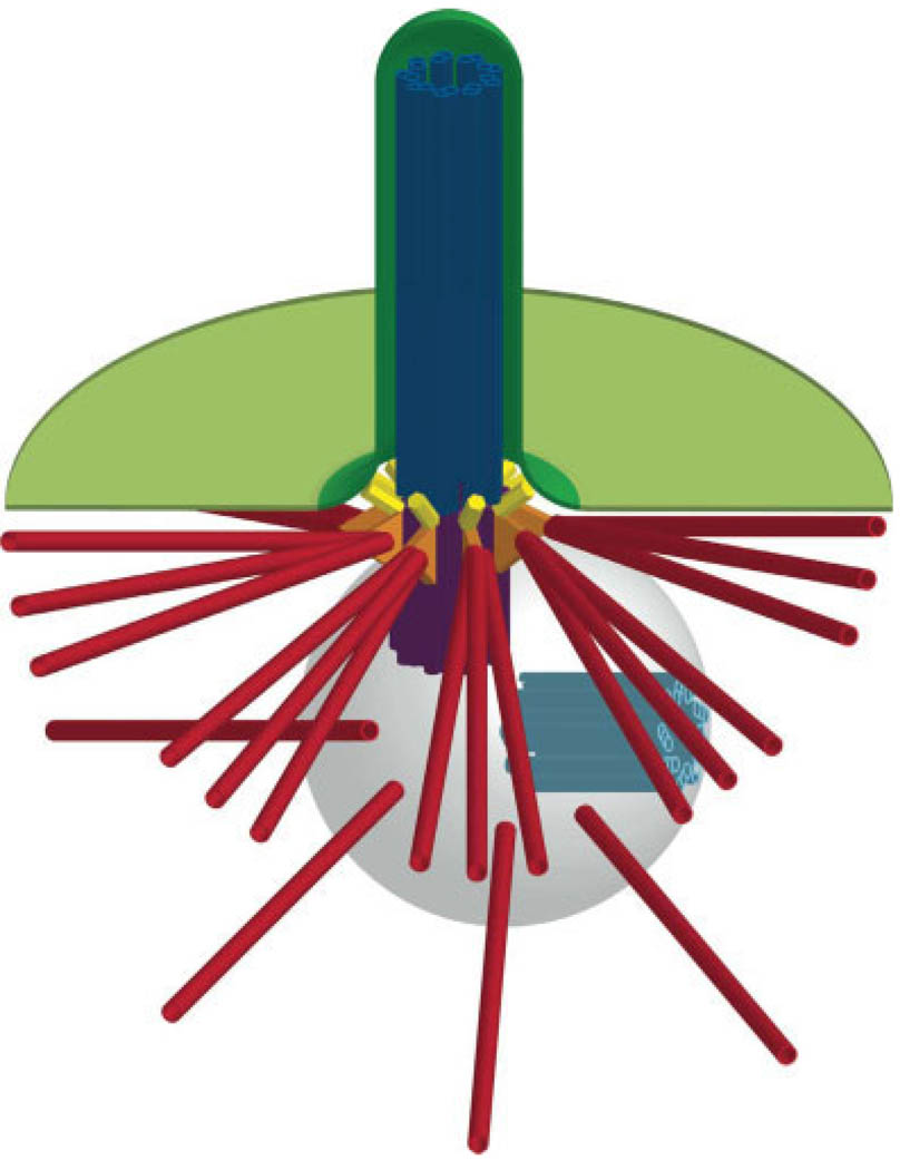
The primary cilium is a solitary, immotile cilium that is present in almost every mammalian cell type. Primary cilia are thought to function as chemosensors, mechanosensors, or both, depending on cell type, and have been linked to several developmental signaling pathways. Primary cilium malfunction has been implicated in several human diseases, the symptoms of which include vision and hearing loss, polydactyly, and polycystic kidneys. Recently, primary cilia have also been implicated in the development and homeostasis of the skeleton. In this review, we discuss the structure and formation of the primary cilium and some of the mechanical and chemical signals to which it could be sensitive, with a focus on skeletal biology. We also raise several unanswered questions regarding the role of primary cilia as mechanosensors and chemosensors and identify potential research avenues to address these questions.
Figures
References
-
- Alieva IB, Vorobjev IA. Vertebrate primary cilia: a sensory part of centrosomal complex in tissue cells, but a “sleeping beauty” in cultured cells? Cell Biol Int. 2004;28:139–150. - PubMed
-
- Bettencourt-Dias M, Glover DM. Centrosome biogenesis and function: centrosomics brings new understanding. Nat Rev Mol Cell Biol. 2007;8:451–463. - PubMed
-
- Brailov I, Bancila M, Brisorgueil MJ, Miquel MC, Hamon M, Verge D. Localization of 5-HT(6) receptors at the plasma membrane of neuronal cilia in the rat brain. Brain Res. 2000;872:271–275. - PubMed
-
- Christensen ST, Pedersen LB, Schneider L, Satir P. Sensory cilia and integration of signal transduction in human health and disease. Traffic. 2007;8:97–109. - PubMed
MeSH terms
Grants and funding
LinkOut - more resources
Full Text Sources
