. 2008 Sep;60(3):640-9.
doi: 10.1002/mrm.21680.

Effects of refocusing flip angle modulation and view ordering in 3D fast spin echo

Affiliations

Effects of refocusing flip angle modulation and view ordering in 3D fast spin echo

Reed F Busse et al. Magn Reson Med. 2008 Sep.

Abstract

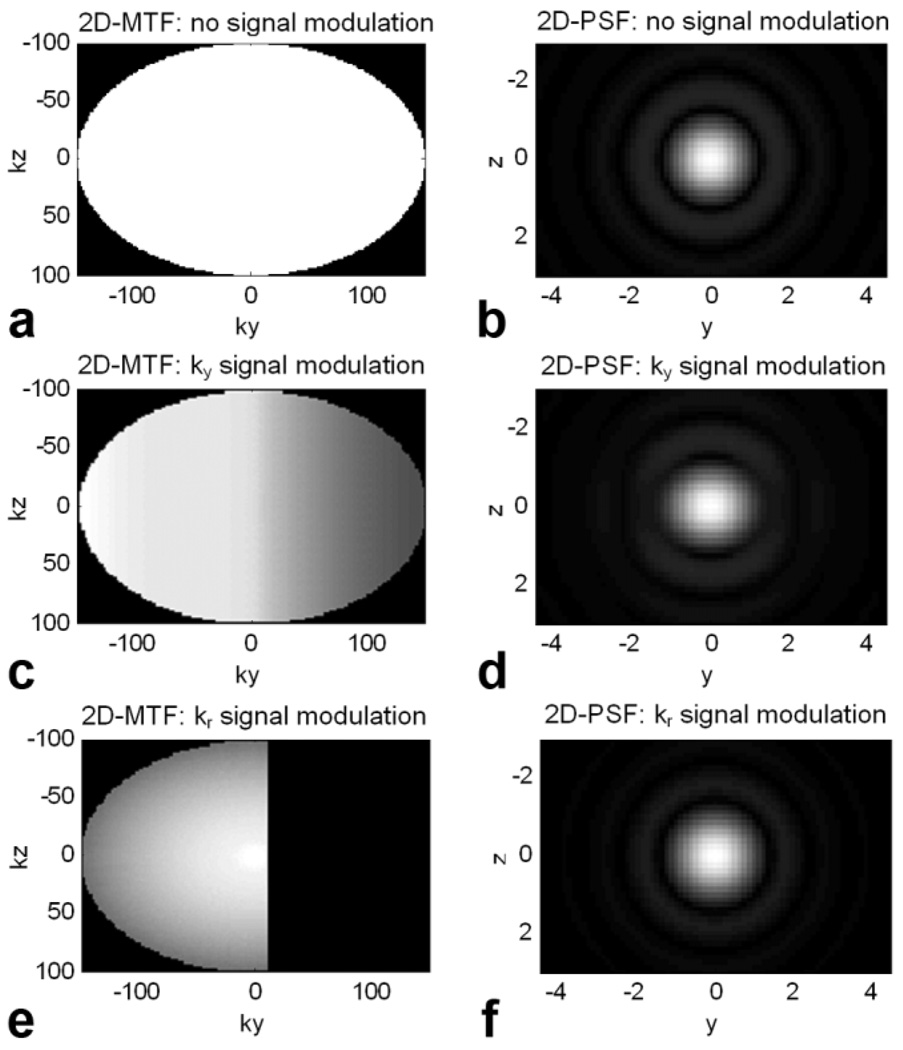
Recent advances have reduced scan time in three-dimensional fast spin echo (3D-FSE) imaging, including very long echo trains through refocusing flip angle (FA) modulation and 2D-accelerated parallel imaging. This work describes a method to modulate refocusing FAs that produces sharp point spread functions (PSFs) from very long echo trains while exercising direct control over minimum, center-k-space, and maximum FAs in order to accommodate the presence of flow and motion, SNR requirements, and RF power limits. Additionally, a new method for ordering views to map signal modulation from the echo train into k(y)-k(z) space that enables nonrectangular k-space grids and autocalibrating 2D-accelerated parallel imaging is presented. With long echo trains and fewer echoes required to encode large matrices, large volumes with high in- and through-plane resolution matrices may be acquired with scan times of 3-6 min, as demonstrated for volumetric brain, knee, and kidney imaging.


Figures

FIG. 1
FIG. 1
Two methods for producing refocusing FA trains with explicit control of αmin, αcent, and αmax. a: For the PSS-signal-envelope method (solid) targets are defined for the PSS signal level (transverse coherence), which increases throughout the train, while for the relaxation-specific method (dashed) targets are for the net signal, which includes relaxation as well as transverse coherence. b: Refocusing FAs that produce these targets are calculated, either ignoring relaxation with the PSS-signal-envelope method (solid), or given a set of T1 and T2 relaxation values with the relaxation-specific method (dashed). The two methods produce similar results given the same αmin, αcent, and αmax values. c: The actual signals in materials with various T1 and T2 values are nearly identical for the two methods. Model parameters: αmin = 25°, αcent = 70°, αmax = 120°, ETL = 100 +6 unacquired echoes at the start, echo spacing = 5 ms; relaxation-specific method: T1 = 1000 ms, T2 = 100 ms.
FIG. 2
FIG. 2
a: A separable sampling grid for 2D acceleration with autocalibration is compatible with previously described 3D-FSE view ordering techniques. b: A more efficient approach, however, is to acquire views on a nonseparable grid, which requires approximately one-third less scan time but requires a more advanced view-ordering method, such as that proposed.
FIG. 3
FIG. 3
The proposed view-ordering algorithm can direct signal modulation along a given direction in ky-kz-space. a: For signal modulation directed along the ky direction, views are sorted first by ky location and assigned echo numbers based on this value —echo numbers are depicted by colors incremented through the spectrum with echoes 1, 15, and 45 circled. b: Next, all views of a given echo number are sorted by kz location and assigned train numbers based on this value—train numbers are depicted by colors incremented through the spectrum with trains 1, 6, 16, and 26 circled, and connecting lines showing view order within these trains. c: Likewise, for signal modulation directed along the kr direction, views are sorted first by kr and assigned echo numbers, then by kθ and assigned train numbers (d).
FIG. 4
FIG. 4
Several effects of changing the minimum refocusing FA, αmin, are demonstrated: (a) FA trains for αmin of 20°, 30°, and 40° (with ETL adjusted to maintain a given desired TEeff = 90 ms) produce (b) different signal modulation curves in a material with T1 = 1000 and T2 = 100 ms; however, the (c) normalized PSFs are very similar. As a function of αmin, (d) ETL is decreased as αmin is increased in order to maintain the same TEeff, making scans longer, but (e) signal loss due to motion also decreases as αmin is increased. Resolution (FWHM of the PSF) is relatively insensitive to αmin given a TEeff-adjusted ETL. Model parameters: αcent = 70°, αmax = 120°, echo spacing = 5 ms.
FIG. 5
FIG. 5
Signal modulation that results from the combined effect of relaxation and FA modulation is mapped into k-space via the specific view order employed. In an ideal situation, (a) no signal modulation would occur, resulting in (b) an optimal PSF. If view order is such that (c) signal modulation occurs in the ky-direction, then (d) the PSF is broadened slightly in the y direction. If view ordering produces (e) radial signal modulation, then (f) the PSF is broadened slightly in both directions. Model parameters: αmin = 25°, αcent = 70°, αmax = 120°, ETL = 100 +4 unacquired echoes at the start, echo spacing = 5 ms, T1 = 1000 ms, T2 = 100 ms.
FIG. 6
FIG. 6
Profiles of the 2D PSF along the y- and z-axes for {T1, T2} values of {1800, 150} (solid black line), {1000, 100} (dashed black line), and {700, 60} ms (dotted black line) and an optimal PSF corresponding to no signal modulation (gray line). For view ordering that produces ky signal modulation, (a) a small amount of point spread increase occurs with decreasing {T1, T2} values in the y direction, but (b) no increase in the z direction. For view ordering that produces kr signal modulation, a small amount of PSF increase is evenly distributed to (c) y and (d) z directions.
FIG. 7
FIG. 7
Whole-brain imaging. a: A T2-weighted image volume is acquired with a 320 × 272 × 192 matrix (0.8 mm superior/inferior [S/I] (frequency) × 0.8 mm anterior/posterior [A/P] (phase) × 0.8 mm right/left [R/L] (slice) acquired voxel size) in 3:49. b: A T2w-FLAIR image volume with a matrix of 256 × 218 × 128 matrix (1.0 mm × 1.0 mm × 1.2 mm acquired voxel size) is acquired in 4:30. Very thin slice acquisitions enable high-resolution coronal and axial reformats, very similar in quality to the native sagittal acquisitions.
FIG. 8
FIG. 8
PD-weighted whole-knee imaging. Radial modulation view ordering produces PD-weighted contrast. A 256 × 256 × 200 (0.6 mm superior/inferior [S/I] (frequency) × 0.6 mm anterior/posterior [A/P] (phase) × 0.7 mm right/left [R/L] (slice) acquired voxel size) matrix is acquired in 4:25 with 2D-accelerated parallel imaging and half-Fourier encoding.
FIG. 9
FIG. 9
Volumetric kidney imaging. A T2-weighted image volume with a 320 × 320 × 128 matrix (1.0 mm superior/inferior [S/I] (frequency) × 1.0 mm right/left [R/L] (phase) × 1.4 mm anterior/posterior [A/P] (slice) acquired voxel size) is acquired in 5:19 with respiratory gating.

References

    1. Hennig J, Nauerth A, Freidburg H. RARE imaging: a fast imaging method for clinical MR. Magn Reson Med. 1986;3:823–833. - PubMed
    1. Listerud J, Einstein S, Outwater E, Kressel HY. First principles of fast spin echo. Magn Reson Q. 1992;8:199–244. - PubMed
    1. Norris DG, Boernert P, Reese T, Leibfritz D. On the application of ultra-fast RARE experiments. Magn Reson Med. 1992;27:142–164. - PubMed
    1. LeRoux P, Hinks RS. Stabilization of echo amplitudes in FSE sequences. Magn Reson Med. 1993;30:183–191. - PubMed
    1. Alsop DC. The sensitivity of low flip angle RARE imaging. Magn Reson Med. 1997;37:176–184. - PubMed