. 2008 Oct;154(1):98-106.
doi: 10.1111/j.1365-2249.2008.03731.x. Epub 2008 Aug 22.

Human TSLP directly enhances expansion of CD8+ T cells

Affiliations

Human TSLP directly enhances expansion of CD8+ T cells

T Akamatsu et al. Clin Exp Immunol. 2008 Oct.

Abstract

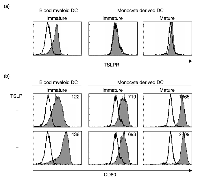
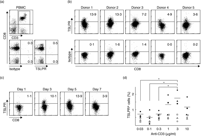
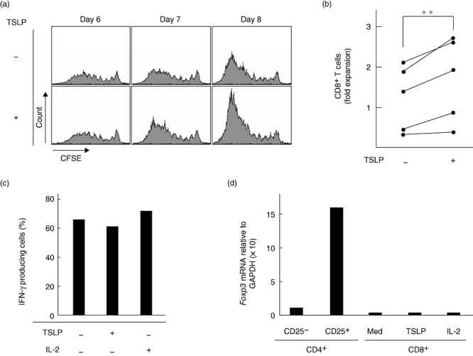
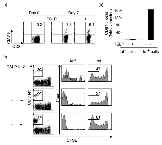
Human thymic stromal lymphopoietin (TSLP) promotes CD4(+) T-cell proliferation both directly and indirectly through dendritic cell (DC) activation. Although human TSLP-activated DCs induce CD8(+) T-cell proliferation, it is not clear whether TSLP acts directly on CD8(+) T cells. In this study, we show that human CD8(+) T cells activated by T-cell receptor stimulation expressed TSLP receptor (TSLPR), and that TSLP directly enhanced proliferation of activated CD8(+) T cells. Although non-stimulated human CD8(+) T cells from peripheral blood did not express TSLPR, CD8(+) T cells activated by anti-CD3 plus anti-CD28 did express TSLPR. After T-cell receptor stimulation, TSLP directly enhanced the expansion of activated CD8(+) T cells. Interestingly, using monocyte-derived DCs pulsed with a cytomegalovirus (CMV)-specific pp65 peptide, we found that although interleukin-2 allowed expansion of both CMV-specific and non-specific CD8(+) T cells, TSLP induced expansion of only CMV-specific CD8(+) T cells. These results suggest that human TSLP directly enhances expansion of CD8(+) T cells and that the direct and indirect action of TSLP on expansion of target antigen-specific CD8(+) T cells may be beneficial to adoptive cell transfer immunotherapy.


Figures

Fig. 4
Fig. 4
TSLPR and CD80 expression on blood myeloid DCs and monocyte-derived DCs. (a) Purified immature CD11c+ blood myeloid DCs and monocyte-derived DCs with or without maturation with IL-6, tumour necrosis factor-α, IL-1β and prostaglandin-E2, were stained with mAb to TSLPR or isotype control. Cell surface marker phenotypes were determined by flow cytometry. Filled histograms represent staining of DCs with TSLPR; open histograms represent isotype controls. Data represent one of three experiments. (b) Indicated DCs were cultured for 24 h with or without TSLP and stained with mAb to CD80 or isotype control. Filled histograms represent staining of DCs with CD80; open histograms represent isotype controls. Numbers in histograms indicate the mean fluorescence intensity. Data represent one of three experiments.
Fig. 1
Fig. 1
TCR stimulation induces TSLPR expression on human CD8+ T cells. (a) PBMCs were stained with mAb to CD3, CD8 and TSLPR or isotype control. Cell surface marker phenotypes were determined by flow cytometry. Lower panels represent staining of CD3+ cells with TSLPR or isotype control. Numbers in quadrants indicate the percentages of cells for each given phenotype in CD3+ cells. Data represent one of five independent experiments. (b, c) Purified human CD8+ T cells were stimulated for 5 days (b) or indicated days (c) with immobilized anti-CD3 plus soluble anti-CD28, and numbers in quadrants indicate the percentages of cells for each given phenotype in CD3+CD8+ cells. Data shown from five individuals (b) and one of five independent experiments (c). (d) CD8+ T cells were stimulated with anti-CD28 and various concentrations of anti-CD3. Data shown are the percentages of TSLPR+ cells in CD3+CD8+ cells in three individual experiments (closed symbols). Horizontal bars indicate the mean of percentages of TSLPR+ cells and open symbols show isotype control from individuals. P-values as determined by the paired t-test. *P < 0·05.
Fig. 2
Fig. 2
CD8+ T cells express TSLPR on their surface in mixed lymphocyte reaction. Purified human CD8+ T cells were cultured with allogeneic MoDCs for 7 days at a DC : T cell ratio of 1:2. Cell surface marker phenotypes were determined by flow cytometry as in Fig. 1b. Data represent one of five independent experiments.
Fig. 3
Fig. 3
TSLP directly enhances expansion of functional CD8+ T cells induced by TCR stimulation. (a) Cell divisions of CFSE-labelled purified human CD8+ T cells stimulated with anti-CD3 plus anti-CD28 with or without TSLP for indicated days. Filled histograms represent CFSE dilution in CD8+ T cells. Data represent one of five individual experiments. (b) Closed circles indicate fold expansion of CD8+ T cells stimulated with anti-CD3 plus anti-CD28 with or without TSLP compared with the initial CD8+ T-cell number in five individual experiments. The lines indicate CD8+ T cells from the same individual. (**P < 0·05). (c) The percentages of IFN-γ producing cells in CD8+ T cells using intracellular cytokine staining. CD8+ T cells were stimulated with anti-CD3 plus anti-CD28 with or without TSLP or IL-2 and restimulated with PMA plus ionomycin. Data represent one of three independent experiments. (d) Expression levels of mRNA encoding Foxp3 were measured using the real-time quantitative RT-PCR. CD8+ T cells were stimulated with anti-CD3 plus anti-CD28 with or without TSLP or IL-2. Data represent one of three independent experiments.
Fig. 5
Fig. 5
Human TSLP induces predominant expansion of CMV-specific CD8+ T cells. (a) Purified human CD8+ T cells were stimulated with autologous MoDCs pulsed with HLA-A24-restricted CMVpp65 peptide for 7 days with or without TSLP at a DC : T cell ratio of 1:2. Cells were stained with mAb to cell surface marker phenotypes and HLA-A2402-CMVpp65 tetramer. (b) Fold expansion of tetramer-CD8+ T cells and tetramer+CD8+ T cells after 7 days of culture with (filled bars) and without (open bars) TSLP. Data represent one of 12 independent experiments. (c) Cell divisions of CFSE-labelled purified human CD8+ T cells stimulated for 7 days with autologous MoDCs pulsed with CMVpp65 peptide (upper panels) or with either TSLP (middle panels) or IL-2 (lower panels). Cells were stained with anti-CD8 and CMVpp65 tetramer. Numbers indicate the percentages of proliferated tetramer+CD8+ T cells (left panels) and proliferated tetramer-CD8+ T cells (right panels) in CD8+ cells. Data represent one of five independent experiments.

References

    1. Reche PA, Soumelis V, Gorman DM, et al. Human thymic stromal lymphopoietin preferentially stimulates myeloid cells. J Immunol. 2001;167:336–43. - PubMed
    1. Soumelis V, Reche PA, Kanzler H, et al. Human epithelial cells trigger dendritic cell-mediated allergic inflammation by producing TSLP. Nat Immunol. 2002;3:673–80. - PubMed
    1. Watanabe N, Hanabuchi S, Soumelis V, et al. Human thymic stromal lymphopoietin promotes dendritic cell-mediated CD4+ T cell homeostatic expantion. Nat Immunol. 2004;5:426–34. - PubMed
    1. Wang YH, Ito T, Wang YH, et al. Maintenance and polarization of human Th2 central memory T cells by thymic stromal lymphopoietin-activated dendritic cells. Immunity. 2006;24:827–38. - PubMed
    1. Gilliet M, Soumelis V, Watanabe N, et al. Human dendritic cells activated by TSLP and CD40L induce proallergic cytotoxic T cells. J Exp Med. 2003;197:1059–63. - PMC - PubMed

Publication types

MeSH terms