. 2008 Nov 14;283(46):31960-7.
doi: 10.1074/jbc.M805431200. Epub 2008 Aug 26.

Modulation of PGC-1 coactivator pathways in brown fat differentiation through LRP130

Affiliations

Modulation of PGC-1 coactivator pathways in brown fat differentiation through LRP130

Marcus P Cooper et al. J Biol Chem. .

Abstract

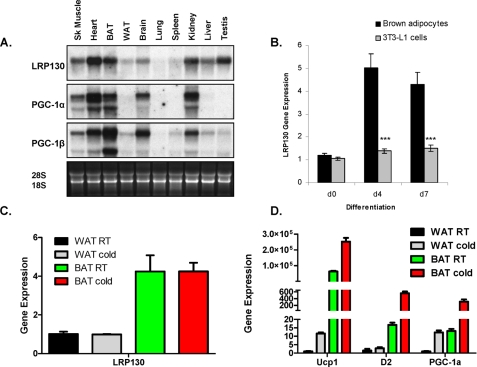
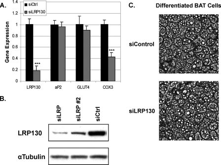
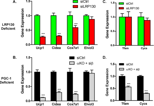
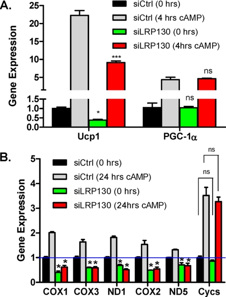
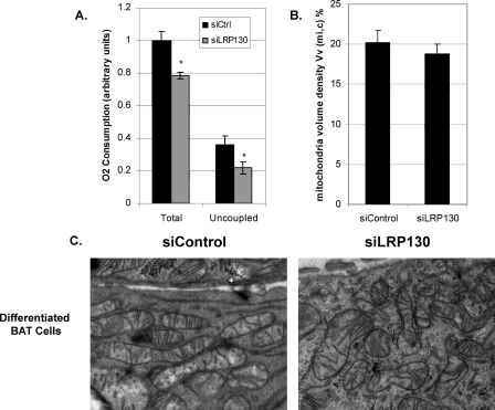
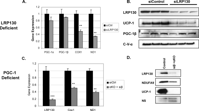
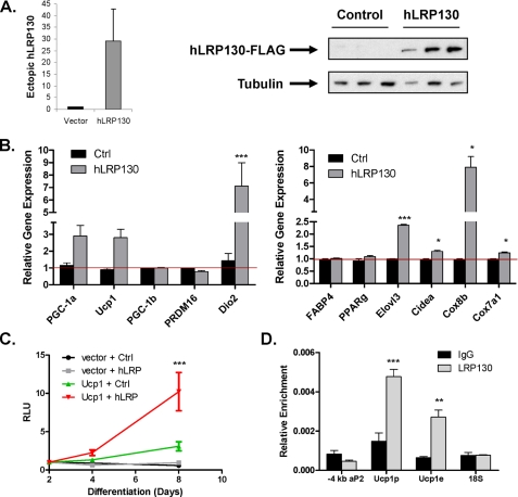
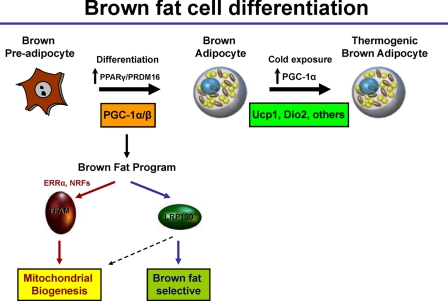
The PGC-1 coactivators are important regulators of oxidative metabolism. We previously demonstrated that LRP130 is a binding partner of PGC-1alpha, required for hepatic gluconeogenesis. LRP130 is the gene mutated in Leigh syndrome French Canadian variant, a rare neurodegenerative disease. The importance of LRP130 in other, non-hepatocyte biology remains obscure. To better understand PGC-1 coactivator function in brown fat development, we explored the metabolic role of LRP130 in brown adipocyte differentiation. We show that LRP130 is preferentially enriched in brown fat compared with white, and induced in a PGC-1-dependent manner during differentiation. Despite intact PGC-1 coactivator expression, brown fat cells deficient for LRP130 exhibit attenuated expression of several genes characteristic of brown fat, including uncoupling protein 1. Oxygen consumption studies support a specific defect in proton leak due to attenuated uncoupling protein 1 expression. Notably, brown fat cell development common to both PGC-1 coactivators is governed by LRP130. Conversely, the cAMP response controlled by PGC-1alpha is not regulated by LRP130. These data implicate LRP130 in brown fat cell development and differentiation.


Figures

FIGURE 1.
FIGURE 1.
Spatiotemporal distribution of LRP130. A, tissue Northern of LRP130, PGC-1α, and PGC-1β. B, gene expression of LRP130 in differentiating brown fat cells versus 3T3 L1 cells. Differentiation mixture was added on day 0 (n = 3). C, gene expression of LRP130; D, select brown fat genes in white and brown fat depots derived from animals housed at room temperature or exposed to cold at 4 °C for 4 h (n = 5). Statistics: Panel D, no significant change in room temperature versus cold in WAT or BAT for LRP130. Panel C, p < 0.05; D2 changes in WAT not significant. Error bars represent mean (±S.E.). *, p < 0.05; **, p < 0.01; ***, p < 0.001.
FIGURE 2.
FIGURE 2.
Adipogenic and morphological features of brown fat cells deficient for LRP130. A, gene expression of select genes at day 7 of differentiation (n = 3). B, protein expression of LRP130 in stably depleted brown fat cells at day 7 differentiation. A detailed gene expression profile for RNAi no. 2 is in supplemental Fig. S1. C, phase contrast of cells stably transduced with control shRNA (siControl) or shRNA against LRP130 (siLRP130). Error bars represent mean (±S.E.). *. p < 0.05; **, p < 0.01; ***, p < 0.001.
FIGURE 3.
FIGURE 3.
LRP130 phenocopies certain features of PGC-1 double-deficient brown fat cells. Designated αKO + siβ, PGC-1-deficient cells are PGC-1α-null cells with stable shPGC-1β knock-down. Designated siCtrl, the control cells contain a control shRNA. Gene expression profile of brown fat genes and mitochondrial genes in LRP130-deficient (A and C) versus PGC-1 double-deficient mature brown fat cells (B and D) (n = 3). In comparison to siCtrl cell, PGC-1α-null cells stably expressing a control shRNA showed no difference (data not shown). Error bars represent mean (±S.E.). *, p < 0.05; **, p < 0.01; ***, p < 0.001.
FIGURE 4.
FIGURE 4.
Depletion of LRP130 does not alter cAMP mediated induction of Ucp1. A, gene expression of Ucp1 and PGC-1α in mature brown fat cells treated with forskolin for 4 h. Untreated siCtrl cells were statistically compared with untreated siLRP130 cells. cAMP-treated siCtrl cells were statistically compared with cAMP-treated siLRP130 cells (n = 4). B, gene expression of several mitochondrial-encoded genes, and nuclear-encoded cytochrome c (Cycs). 24 h of cAMP treatment proved optimal for inducing these genes (n = 4). Untreated siCtrl cells were statistically compared with untreated siLRP130 cells. cAMP-treated siCtrl cells were statistically compared with cAMP-treated siLRP130 cells. Note, except for ND5 all mitochondrial genes in the siCtrl cells were significantly induced (p < 0.05) following cAMP treatment. Statistical analysis: Two-way analysis of variance with Bonferroni post-test. Error bars represent mean (±S.E.). *, p < 0.05; **, p < 0.01; ***, p < 0.001.
FIGURE 5.
FIGURE 5.
LRP130-deficient brown fat cells have a specific block in proton leak. A, total respiration and proton leak (“uncoupled”) in intact mature brown fat cells (n = 6). B, mitochondrial volume density and C, electron micrographs of mitochondria from stable shControl (siControl) or shLRP130 (siLRP130) mature brown fat cells. Error bars represent mean (±S.E.). *, p < 0.05; **, p < 0.01; ***, p < 0.001.
FIGURE 6.
FIGURE 6.
The PGC-1 coactivators regulate LRP130 gene expression. Expression of the PGC-1 coactivators, Ucp1 and some mitochondrial genes in LRP130-deficient cells. Left, mRNA (n = 3). Right, protein. (A, mRNA and B, protein) versus LRP130 and select mitochondrial gene expression in PGC-1 double-deficient cells (C, mRNA and D, protein). DesignatedαKO + siβ, PGC-1-deficient cells are PGC-1α-null cells with stable shPGC-1β knock-down. Designated siCtrl, control cells are wild-type cells stably expression a control shRNA. In comparison to siCtrl cell, PGC-1α-null cells stably expressing a control shRNA showed no difference (data not shown). In cells null for PGC-1α or deficient in PGC-1β, LPR130 gene expression was not affected (supplemental Fig. S2). Error bars represent mean (±S.E.). *, p < 0.05; **, p < 0.01; ***, p < 0.001.
FIGURE 7.
FIGURE 7.
LRP130 induces Ucp1 and is enriched on the Ucp1 promoter. A, stable expression of exogenous human LRP130-FLAG in mature 3T3-F442A cells (n = 3). B, expression of brown fat genes in mature 3T3-F442A cells, stably expressing vector or human LRP130-FLAG (n = 3). C, luciferase reporter assay using the -4kbto 1+ bp Ucp1 promoter in mature 3T3-F442A cells. Measurements were performed on days 2, 4, and 8 of differentiation. RLU denotes relative luciferase units (n = 3). D, chromatin immunoprecipitation of LRP130 in mature brown fat cells. Q-PCR was used to quantify enrichment at the -4 kb PPRE of the aP2 promoter (-4kb aP2), + 1 to -200 bp region of the Ucp1 promoter (Ucp1 p), -2500 to -2300 bp region of the Ucp1 enhancer(Ucp1 e), and a random control site in the 18S gene. Error bars represent mean (±S.E.). *, p < 0.05; **, p < 0.01; ***, p < 0.001.
FIGURE 8.
FIGURE 8.
Model depicting the role of LRP130 in brown fat cell differentiation and function. PPARγ and PRDM16 are important upstream regulators, induced after differentiation. Subsequently, the PGC-1 coactivators are induced to drive two programs, mitochondrial biogenesis and uncoupled respiration. The PGC-1s regulate ERRα and NRFs to induce TFAM and other factors critical for mitochondrial biogenesis. The PGC-1s induce LRP130, an induction important for remodeling of mitochondria. Inside the mitochondrion (dotted arrow), LRP130 regulates mitochondrial-encoded gene expression. Inside the nucleus it regulates Ucp1 expression, as well as, certain brown fat genes downstream of the PGC-1 coactivators. LPR130 may amplify the process by inducing PGC-1α in an autoregulatory loop. After basal Ucp1 expression is established, PGC-1α governs the cold response essential for thermogenesis.

References

    1. Hansen, J. B., and Kristiansen, K. (2006) Biochem. J. 398 153-168 - PMC - PubMed
    1. Cannon, B., and Nedergaard, J. (2004) Physiol. Rev. 84 277-359 - PubMed
    1. Hull, D., and Segall, M. M. (1965) J. Physiol 181 468-477 - PMC - PubMed
    1. Simon, H. E. (1950) Am. J. Surg. 80 127-130 - PubMed
    1. Dawkins, M. J., and Hull, D. (1964) J. Physiol. 172 216-238 - PMC - PubMed

Publication types

MeSH terms