. 2008 Nov 21;283(47):32412-8.
doi: 10.1074/jbc.M805918200. Epub 2008 Aug 25.

A thermodynamic model for Nap1-histone interactions

Affiliations

A thermodynamic model for Nap1-histone interactions

Andrew J Andrews et al. J Biol Chem. .

Abstract

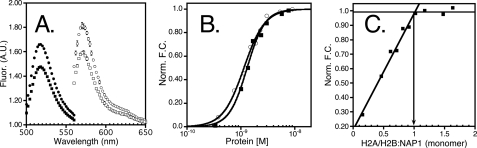
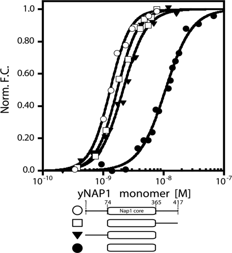
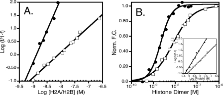
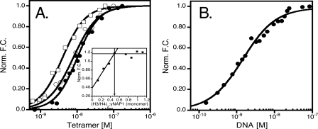
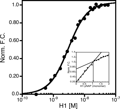
The yeast nucleosome assembly protein 1 (yNap1) plays a role in chromatin maintenance by facilitating histone exchange as well as nucleosome assembly and disassembly. It has been suggested that yNap1 carries out these functions by regulating the concentration of free histones. Therefore, a quantitative understanding of yNap1-histone interactions also provides information on the thermodynamics of chromatin. We have developed quantitative methods to study the affinity of yNap1 for histones. We show that yNap1 binds H2A/H2B and H3/H4 histone complexes with low nm affinity, and that each yNap1 dimer binds two histone fold dimers. The yNap1 tails contribute synergistically to histone binding while the histone tails have a slightly repressive effect on binding. The (H3/H4)(2) tetramer binds DNA with higher affinity than it binds yNap1.


Figures

FIGURE 1.
FIGURE 1.
Fluorescence change as a function of protein binding. A, fluorescence (A.U.) as a function of wavelength (nm), with closed circles/squares representing the signal from the excitation of Alexa-488 (yNap1m*) in the presence (closed squares) or absence (closed circles) of H2A/H2B and the open circles/squares representing Alexa-546 (H2A/H2B(T112C)) in the presence (open squares) or absence (open circles) of yNap1. B, normalized fluorescence change as a function of either H2A/H2B titrated into Alexa-546-labeled yNap1 (closed squares) or yNap1 titrated into Alexa-546 labeled H2A/H2B (open circles). C, change in fluorescence plotted as a function of the ratio of H2A/H2B to yNap1. The intersection of the linear phase with the plateau is equal to the molar ratio at which yNap1 is saturated. These data suggest that two H2A/H2B dimers bind one yNap1 dimer or that one H2A/H2B binds one yNap1 monomer. The conditions were 20–50 mm Tris, pH 7.5, 1 mm dithiothreitol, 0.1 mg/ml bovine serum albumin, 90–120 mm NaCl, (1–5) × 10–10 m Alexa-labeled protein, and (0–5) × 10–7 m non-labeled protein. For results of the fit see Table 1.
FIGURE 2.
FIGURE 2.
Affinity and stoichiometry of the yNap1-H2A/H2B dimer complex. Normalized change in fluorescence as a function of yNap1 (open circles), yNap1(74–417) (open squares), yNap1(1–365) (closed triangles), and yNap1(74–365) (closed circles), all as monomers. The conditions were 20–50 mm Tris, pH 7.5, 1 mm dithiothreitol, 0.1 mg/ml bovine serum albumin, 90–120 mm NaCl, (0.5–1) × 10–10 m Alexa-488-H2A/H2B(T112C), and (0–5 × 10⁻⁷ m yNap1 or its truncations. For results of the fit see Table 1.
FIGURE 3.
FIGURE 3.
Cooperativity is dependent on both the ionic strength and histone tails. A, Hill plot of H2A/H2B binding yNap1 at ∼0.15 m (closed circles) and ∼0.35 m (open squares-same data in all plots) ionic strength. The slopes of these data result in a nH of 1 ± 0.1 and 2.4 ± 0.2, respectively. B, normalized fluorescence change as a function of histone dimer. Closed circles are tail-less dimers, and open squares are major type histone dimer. Inset of B is the Hill plot of the tail-less (closed circles) and major type (open squares) dimer. The slopes of these data result in an nH of 1.5 ± 0.1 and 1.0 ± 0.1, respectively.
FIGURE 4.
FIGURE 4.
The interaction of yNap1 and DNA with histone tetramer. A, normalized fluorescence change as a function of histone tetramer binding to (yNap1m*)-Alexa-546 (closed circles), tail-less (H3/H4)2 (open squares), and H3(H113A)/H4 (plotted as tetramer) (open squares). Inset of A is the stoichiometry of (H3/H4)2 to yNAP dimer. (H3/H4)2 was titrated against 1×10⁻⁷ m (yNap1m*)-Alexa-546 to result in a stoichiometry of 1 histone tetramer to 1 yNap1 dimer. B, normalized fluorescence change as a function of DNA binding to (H3/H4(E63C))2-Alexa-488 (10–10 m). Buffer conditions were 20–50 mm Tris, pH 7.5, 1 mm dithiothreitol, 0.1 mg/ml bovine serum albumin, and 300 mm NaCl.
FIGURE 5.
FIGURE 5.
Binding and stoichiometry of H1 to yNap1. The normalized fluorescence change as a function of linker histone H1 binding to (yNap1m*)-Alexa-546. Inset is the stoichiometry of H1 to yNap1. While two phases were easily discernible the second did not level out as expected. These data suggest specific binding of two H1 linker histones to yNap1 and a possible third nonspecific H1-yNap1 interaction.

References

    1. De Koning, L., Corpet, A., Haber, J. E., and Almouzni, G. (2007) Nat. Struct. Mol. Biol. 14 997–1007 - PubMed
    1. Eitoku, M., Sato, L., Senda, T., and Horikoshi, M. (2008) Cell Mol. Life Sci. 65 414–444 - PMC - PubMed
    1. Park, Y. J., and Luger, K. (2006) Biochem. Cell Biol. 84 549–558 - PubMed
    1. Zlatanova, J., Seebart, C., and Tomschik, M. (2007) Faseb J. 21 1294–1310 - PubMed
    1. Park, Y. J., and Luger, K. (2006) Proc. Natl. Acad. Sci. U. S. A. 103 1248–1253 - PMC - PubMed

Publication types

MeSH terms