. 2008 Sep 2;105(35):12991-6.
doi: 10.1073/pnas.0805802105. Epub 2008 Aug 26.

Immunosuppressive therapy mitigates immunological rejection of human embryonic stem cell xenografts

Affiliations

Immunosuppressive therapy mitigates immunological rejection of human embryonic stem cell xenografts

Rutger-Jan Swijnenburg et al. Proc Natl Acad Sci U S A. .

Abstract

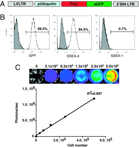
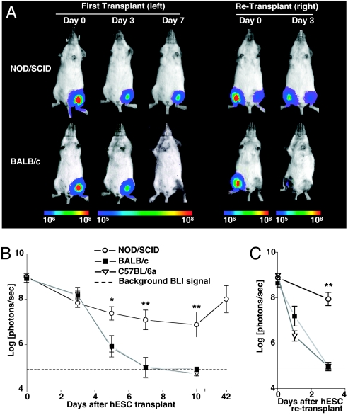
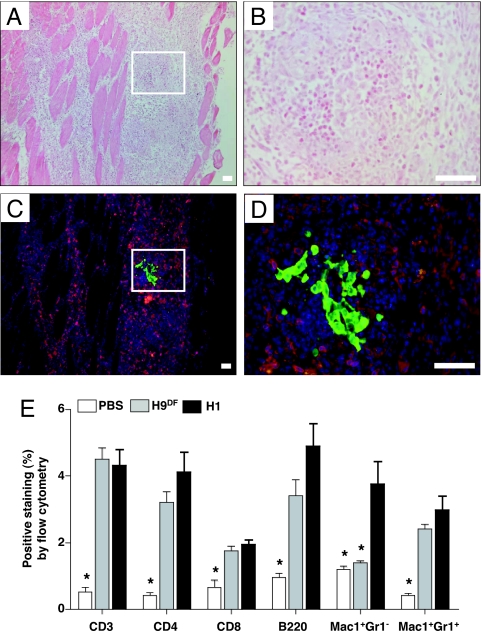
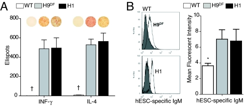
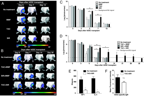
Given their self-renewing and pluripotent capabilities, human embryonic stem cells (hESCs) are well poised as a cellular source for tissue regeneration therapy. However, the host immune response against transplanted hESCs is not well characterized. In fact, controversy remains as to whether hESCs have immune-privileged properties. To address this issue, we used in vivo bioluminescent imaging to track the fate of transplanted hESCs stably transduced with a double-fusion reporter gene consisting of firefly luciferase and enhanced GFP. We show that survival after transplant is significantly limited in immunocompetent as opposed to immunodeficient mice. Repeated transplantation of hESCs into immunocompetent hosts results in accelerated hESC death, suggesting an adaptive donor-specific immune response. Our data demonstrate that transplanted hESCs trigger robust cellular and humoral immune responses, resulting in intragraft infiltration of inflammatory cells and subsequent hESC rejection. Moreover, we have found CD4(+) T cells to be an important modulator of hESC immune-mediated rejection. Finally, we show that immunosuppressive drug regimens can mitigate the anti-hESC immune response and that a regimen of combined tacrolimus and sirolimus therapies significantly prolongs survival of hESCs for up to 28 days. Taken together, these data suggest that hESCs are immunogenic, trigger both cellular and humoral-mediated pathways, and, as a result, are rapidly rejected in xenogeneic hosts. This process can be mitigated by a combined immunosuppressive regimen as assessed by molecular imaging approaches.


Conflict of interest statement

The authors declare no conflict of interest.

Figures

Fig. 1.
Fig. 1.
Characterization of the DF fLuc and enhanced eGFP transduced hESCs. (A) Schema of the DF reporter gene containing fLuc and eGFP driven by a human ubiquitin promoter. (B) Flow cytometric analysis of H9DF hESCs shows robust expression of eGFP. Transduced hESCs are largely positive for SSEA-4, and negative for SSEA-1, confirming their pluripotent state. (C) Stably transduced hESCs show robust correlation between cell number and reporter gene activity. BLI of a 24-well plate containing increasing numbers of H9DF hESCs are shown above the corresponding graph depicting correlation between cell number and fLuc activity.
Fig. 2.
Fig. 2.
In vivo visualization of hESC survival. (A) Representative BLI images of H9DF hESC transplanted animals show a rapid decrease in BLI signal in immunocompetent animals (BALB/c), as opposed to immunodeficient (NOD/SCID) mice, reaching background levels at day 7 after transplant. Accelerated BLI signal loss in BALB/c animals was seen after repeated hESC transplantation into the contralateral gastrocnemius muscle. Color scale bar values are in photons per second per square centimeter per steradian (sr). (B and C) Graphical representation of longitudinal BLI after primary (B) and secondary (C) hESC transplantation into immunodeficient (NOD/SCID, n = 5) and two immunocompetent (BALB/c and C57BL/6a, n = 5 per group) mouse strains. Note that in NOD/SCID animals, starting at the 10th day after transplant, BLI intensity increases progressively, suggesting hESC proliferation. *, P < 0.05, **, P < 0.01.
Fig. 3.
Fig. 3.
Robust inflammatory cell infiltration after intramuscular hESC transplantation. (A and B) Histopathological evaluation by H&E staining of muscle sections of BALB/c animals, obtained at 5 days after H9DF hESC transplantation, demonstrates robust intramuscular inflammatory cell infiltration at low power (A) and high power (B) view. (C and D) Immunofluorescent staining on corresponding sections reveals abundant presence of CD3+ T cells (red) surrounding eGFP+ hESCs (green). Counterstaining was performed with 4,6-diamidino-2-phenylindole (DAPI, blue). (Scale bars, 50 μm.) (E) FACS analysis of enzymatically digested muscles revealed intra- H9DF and H1 hESC graft infiltration of CD3+ T cells, CD4+ Th cells, CD8+ cytotoxic T cells, B220+ B cells, and Mac-1+Gr-1+ neutrophils at significantly higher intensities compared with PBS injections. Mac-1+Gr-1 (macrophages) cells had a significantly higher presence only in the H1 group. *, P < 0.05.
Fig. 4.
Fig. 4.
hESC transplantation triggers cellular and humoral murine immune responses. (A) ELISPOT assay revealed significantly higher production of both INF-γ and IL-4 by both H1 and H9DF hESC recipient BALB/c splenocytes (n = 6) compared with wild-type animals (n = 3). Representative images of ELISPOT wells are shown above the corresponding bars. †P < 0.001 (B) Representative flow cytometry histograms (Left) and graphical representation (Right) of hESC-specific xeno-reactive IgM antibodies detected at significantly higher rate in H1 and H9DF hESC recipient BALB/c sera (n = 6) compared with WT animals (n = 3). *, P < 0.05.
Fig. 5.
Fig. 5.
Immunosuppressive drug treatment prolongs survival of transplanted hESCs and mitigates adaptive immune response. (A and B) Representative BLI images of H9DF hESCs-transplanted mice receiving no treatment compared with those receiving immunosuppressive monotherapy (MMF, TAC, or SIR) (A) or combined therapy (TAC+MMF, SIR+MMF, TAC+SIR) (B). Although SIR as monotherapy extended hESC survival significantly, TAC+SIR combination therapy proved to be optimal and extended survival of the cells up to day 28 after transplant. Color scale bar values are in photons per second per squared centimeter per sr. (C and D) Graphical representation of single (C) or combined (D) drug treatment efficacy on hESC survival after transplant (n = 5 per group). *, P < 0.05, **, P < 0.01. (E and F) Combined TAC+SIR treatment effectively suppressed INF-γ and IL-4 production by hESC recipient splenocytes (**, P < 0.01) (E) and reduced production of donor-specific xeno-reactive antibodies (F) (P = 0.14; n.s. = not significant).

References

    1. Thomson JA, et al. Embryonic stem cell lines derived from human blastocysts. Science. 1998;282:1145–1147. - PubMed
    1. Laflamme MA, et al. Cardiomyocytes derived from human embryonic stem cells in pro-survival factors enhance function of infarcted rat hearts. Nat Biotechnol. 2007;25:1015–1024. - PubMed
    1. Ben-Hur T, et al. Transplantation of human embryonic stem cell-derived neural progenitors improves behavioral deficit in Parkinsonian rats. Stem Cells. 2004;22:1246–1255. - PubMed
    1. Swijnenburg RJ, van der Bogt KE, Sheikh AY, Cao F, Wu JC. Clinical hurdles for the transplantation of cardiomyocytes derived from human embryonic stem cells: Role of molecular imaging. Curr Opin Biotechnol. 2007;18:38–45. - PubMed
    1. Bradley JA, Bolton EM, Pedersen RA. Stem cell medicine encounters the immune system. Nat Rev Immunol. 2002;2:859–871. - PubMed

Publication types

MeSH terms

Substances